Synthesized X-Ray Images Help Train AI Programs
|
By HospiMedica International staff writers Posted on 17 Jul 2018 |
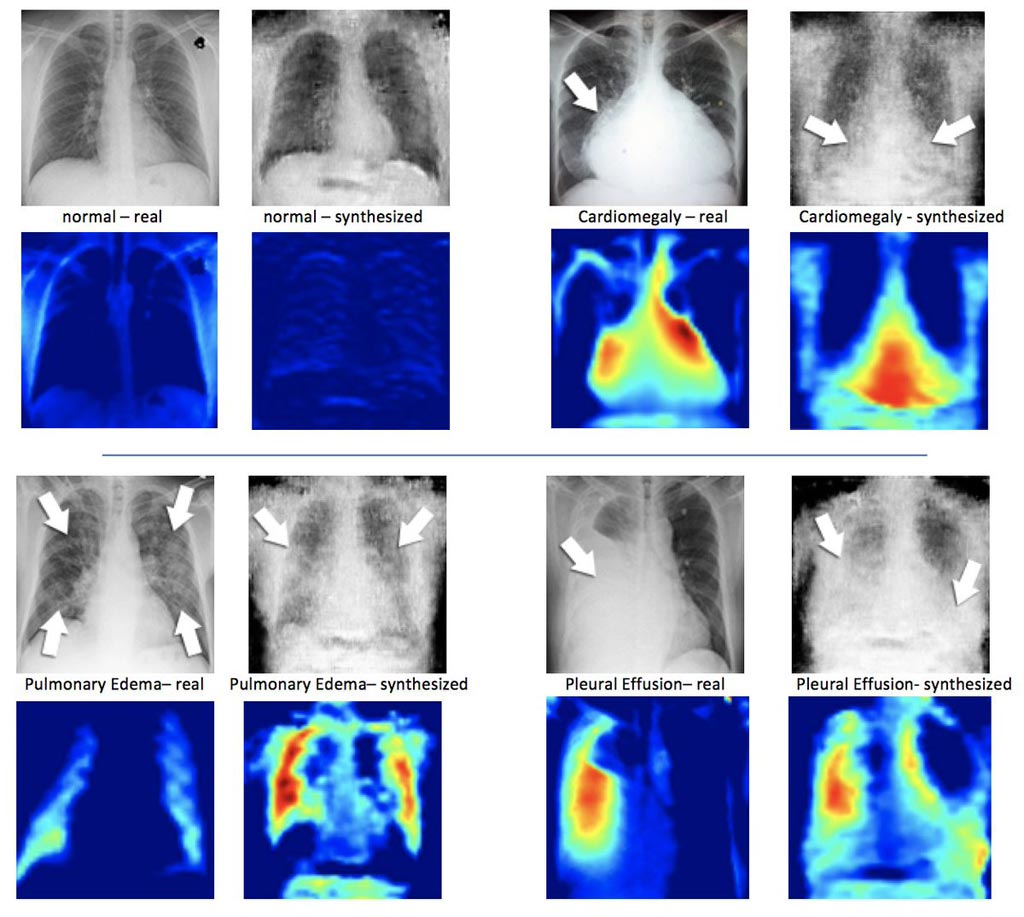
Image: Real X-ray image (L) next to a synthesized X-ray created by DCGAN. Underneath the X-ray images are the corresponding heatmaps (Photo courtesy of Hojjat Salehinejad/MIMLab).
A new study describes how computer generated X-rays can be used to augment artificial intelligence (AI) training sets.
In order to generate and continually improve artificial X-rays, researchers at the University of Toronto (Canada) used deep convolutional generative adversarial network (DCGAN) algorithms, which are made up of two networks: one that generates the images, and the other that tries to discriminate synthetic images from real images. The two networks are continuously trained until they reach a point in which the discriminator cannot differentiate real images from synthesized ones. Once a sufficient number of artificial X-rays are created, they are used to train another DCGAN that can classify the images accordingly.
The researchers then compared the accuracy of the artificially augmented dataset to the original one when fed through their AI system, and found that classification accuracy improved by 20% for common conditions. For some rare conditions, accuracy improved up to 40%. An advantage of the method is that as the synthetic X-rays are not real, the dataset can be readily available to researchers outside hospital premises without violating privacy concerns. The study was presented at the IEEE International Conference on Acoustics, Speech and Signal Processing, held during April 2018 in Calgary (Canada).
“In a sense, we are using machine learning to do machine learning,” said senior author and study presenter Professor Shahrokh Valaee, PhD, of the Machine Intelligence in Medicine Lab (MIMLab). “We are creating simulated X-rays that reflect certain rare conditions so that we can combine them with real X-rays to have a sufficiently large database to train the neural networks to identify these conditions in other X-rays.”
“Deep learning only works if the volume of training data is large enough, and this is one way to ensure we have neural networks that can classify images with high precision,” concluded Professor Valaee. “We've been able to show that artificial data generated by deep convolutional GANs can be used to augment real datasets. This provides a greater quantity of data for training and improves the performance of these systems in identifying rare conditions.”
Related Links:
University of Toronto
Channels
Critical Care
view channel
Light-Based Technology to Measure Brain Blood Flow Could Diagnose Stroke and TBI
Monitoring blood flow in the brain is crucial for diagnosing and treating neurological conditions such as stroke, traumatic brain injury (TBI), and vascular dementia. However, current imaging methods like... Read more
AI Heart Attack Risk Assessment Tool Outperforms Existing Methods
For decades, doctors have relied on standardized scoring systems to assess patients with the most common type of heart attack—non-ST-elevation acute coronary syndrome (NSTE-ACS). The GRACE score, used... Read moreSurgical Techniques
view channel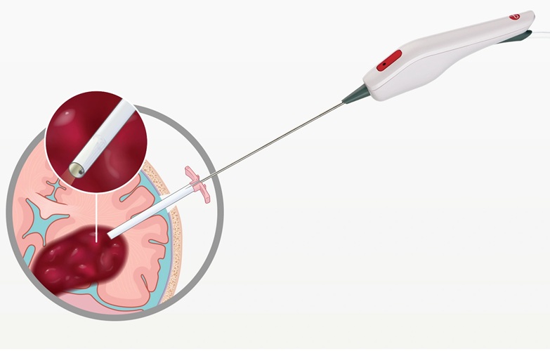
Minimally Invasive Endoscopic Surgery Improves Severe Stroke Outcomes
Intracerebral hemorrhage, a type of stroke caused by bleeding deep within the brain, remains one of the most challenging neurological emergencies to treat. Accounting for about 15% of all strokes, it carries... Read more
Novel Glue Prevents Complications After Breast Cancer Surgery
Seroma and prolonged lymphorrhea are among the most common complications following axillary lymphadenectomy in breast cancer patients. These postoperative issues can delay recovery and postpone the start... Read morePatient Care
view channel
Revolutionary Automatic IV-Line Flushing Device to Enhance Infusion Care
More than 80% of in-hospital patients receive intravenous (IV) therapy. Every dose of IV medicine delivered in a small volume (<250 mL) infusion bag should be followed by subsequent flushing to ensure... Read more
VR Training Tool Combats Contamination of Portable Medical Equipment
Healthcare-associated infections (HAIs) impact one in every 31 patients, cause nearly 100,000 deaths each year, and cost USD 28.4 billion in direct medical expenses. Notably, up to 75% of these infections... Read more
Portable Biosensor Platform to Reduce Hospital-Acquired Infections
Approximately 4 million patients in the European Union acquire healthcare-associated infections (HAIs) or nosocomial infections each year, with around 37,000 deaths directly resulting from these infections,... Read moreFirst-Of-Its-Kind Portable Germicidal Light Technology Disinfects High-Touch Clinical Surfaces in Seconds
Reducing healthcare-acquired infections (HAIs) remains a pressing issue within global healthcare systems. In the United States alone, 1.7 million patients contract HAIs annually, leading to approximately... Read moreHealth IT
view channel
Printable Molecule-Selective Nanoparticles Enable Mass Production of Wearable Biosensors
The future of medicine is likely to focus on the personalization of healthcare—understanding exactly what an individual requires and delivering the appropriate combination of nutrients, metabolites, and... Read moreBusiness
view channel
Philips and Masimo Partner to Advance Patient Monitoring Measurement Technologies
Royal Philips (Amsterdam, Netherlands) and Masimo (Irvine, California, USA) have renewed their multi-year strategic collaboration, combining Philips’ expertise in patient monitoring with Masimo’s noninvasive... Read more
B. Braun Acquires Digital Microsurgery Company True Digital Surgery
The high-end microsurgery market in neurosurgery, spine, and ENT is undergoing a significant transformation. Traditional analog microscopes are giving way to digital exoscopes, which provide improved visualization,... Read more
CMEF 2025 to Promote Holistic and High-Quality Development of Medical and Health Industry
The 92nd China International Medical Equipment Fair (CMEF 2025) Autumn Exhibition is scheduled to be held from September 26 to 29 at the China Import and Export Fair Complex (Canton Fair Complex) in Guangzhou.... Read more


