Real-Time Imaging Shows How SARS-CoV-2 Attacks Human Cells
|
By HospiMedica International staff writers Posted on 10 Sep 2020 |
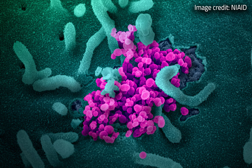
Image: Real-Time Imaging Shows How SARS-CoV-2 Attacks Human Cells (Photo courtesy of NIAID)
Scientists have used imaging assays to view the binding of the spike, or protein, on the SARS-CoV-2 virus to ACE 2 (angiotensin converting enzyme 2) and subsequent internalization that takes place when ACE2 and the spike protein interact in real time.
A report by Science X on Phys.org explains how scientists from the NIH’s National Center for Advancing Translational Sciences (NCATS Bethesda, MA, USA) performed real-time imaging to show how SARS-CoV-2 attacks human cells. Researchers around the world have been trying to understand how SARS-CoV-2 interacts with and penetrates host cells in order to block the stage and halt the onset of COVID19. The SARS-CoV-2 virus uses spike proteins to mobilize its viral DNA into a host cell while the ACE2 receptors, which are human cell proteins, open the door for the attack. In previous studies, researchers had been able to tag these receptor proteins with a green fluorescent protein to image their movements. However, details about the spike protein interactions have been gathered mainly from biochemical or proximity assays and tests with proteins and parts of proteins taken from the virus - "pseudo-viro-particles" according to the Phys.org report. As a result of the lack of fluorescent labeling of these viral proteins, researchers have been unable to perform imaging of their role in the ACE2 receptor binding and subsequent internalization - endocytosis.
Scientists at NCATS who had already begun working on various imaging assays for cancers, viruses and lysosomal storage diseases, used their expertise with nanoparticles for cellular delivery and biosensing to aid in the search for COVID-19 drug treatments. The team started looking at potential ways of applying the protein-nanostructure conjugation techniques. With two proteins that share a binding affinity - a quantum dot attached to one and a fluorescing nanoparticle attached to the other - binding between the two proteins brought the nanostructures close enough for energy transfer between them. The resulting fluorescence quenching allowed the scientists to monitor the protein binding, states the Phys.org report.
The scientists then went on to develop a "pseudovirion" with the potent parts of SARS-CoV-2 spike proteins (where the receptor binding domain is situated) attached to the quantum dot in such a way that the spike proteins continue to attack and penetrate cells similar to an active virus. In order to watch the pseudovirion interacting with ACE2 in a real cell, the quantum dot on the pseudovirion was required to be engineered for emitting at a wavelength that was simple to distinguish from the green fluorescent protein on ACE2, as against optimizing nanoparticle quenching. Using the two clear signals, the NCATS scientists could track the binding of the two proteins and subsequent endocytosis. The team could also see that the binding and endocytosis was halted in the presence of two test antibodies.
"What we're doing here is actually visualizing binding of the spike to ACE 2 (angiotensin converting enzyme 2)," Kirill Gorshkov a research scientist at NCATS told Phys.org. "You can actually see that happen in real time. That's the beauty of this assay and that's why we think it will be important for drug screening."
Related Links:
National Center for Advancing Translational Sciences
Latest COVID-19 News
- Low-Cost System Detects SARS-CoV-2 Virus in Hospital Air Using High-Tech Bubbles
- World's First Inhalable COVID-19 Vaccine Approved in China
- COVID-19 Vaccine Patch Fights SARS-CoV-2 Variants Better than Needles
- Blood Viscosity Testing Can Predict Risk of Death in Hospitalized COVID-19 Patients
- ‘Covid Computer’ Uses AI to Detect COVID-19 from Chest CT Scans
- MRI Lung-Imaging Technique Shows Cause of Long-COVID Symptoms
- Chest CT Scans of COVID-19 Patients Could Help Distinguish Between SARS-CoV-2 Variants
- Specialized MRI Detects Lung Abnormalities in Non-Hospitalized Long COVID Patients
- AI Algorithm Identifies Hospitalized Patients at Highest Risk of Dying From COVID-19
- Sweat Sensor Detects Key Biomarkers That Provide Early Warning of COVID-19 and Flu
- Study Assesses Impact of COVID-19 on Ventilation/Perfusion Scintigraphy
- CT Imaging Study Finds Vaccination Reduces Risk of COVID-19 Associated Pulmonary Embolism
- Third Day in Hospital a ‘Tipping Point’ in Severity of COVID-19 Pneumonia
- Longer Interval Between COVID-19 Vaccines Generates Up to Nine Times as Many Antibodies
- AI Model for Monitoring COVID-19 Predicts Mortality Within First 30 Days of Admission
- AI Predicts COVID Prognosis at Near-Expert Level Based Off CT Scans
Channels
Critical Care
view channel
Light-Based Technology to Measure Brain Blood Flow Could Diagnose Stroke and TBI
Monitoring blood flow in the brain is crucial for diagnosing and treating neurological conditions such as stroke, traumatic brain injury (TBI), and vascular dementia. However, current imaging methods like... Read more
AI Heart Attack Risk Assessment Tool Outperforms Existing Methods
For decades, doctors have relied on standardized scoring systems to assess patients with the most common type of heart attack—non-ST-elevation acute coronary syndrome (NSTE-ACS). The GRACE score, used... Read moreSurgical Techniques
view channel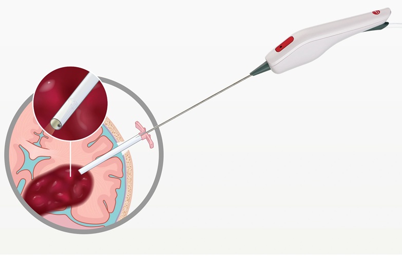
Minimally Invasive Endoscopic Surgery Improves Severe Stroke Outcomes
Intracerebral hemorrhage, a type of stroke caused by bleeding deep within the brain, remains one of the most challenging neurological emergencies to treat. Accounting for about 15% of all strokes, it carries... Read more
Novel Glue Prevents Complications After Breast Cancer Surgery
Seroma and prolonged lymphorrhea are among the most common complications following axillary lymphadenectomy in breast cancer patients. These postoperative issues can delay recovery and postpone the start... Read morePatient Care
view channel
Revolutionary Automatic IV-Line Flushing Device to Enhance Infusion Care
More than 80% of in-hospital patients receive intravenous (IV) therapy. Every dose of IV medicine delivered in a small volume (<250 mL) infusion bag should be followed by subsequent flushing to ensure... Read more
VR Training Tool Combats Contamination of Portable Medical Equipment
Healthcare-associated infections (HAIs) impact one in every 31 patients, cause nearly 100,000 deaths each year, and cost USD 28.4 billion in direct medical expenses. Notably, up to 75% of these infections... Read more
Portable Biosensor Platform to Reduce Hospital-Acquired Infections
Approximately 4 million patients in the European Union acquire healthcare-associated infections (HAIs) or nosocomial infections each year, with around 37,000 deaths directly resulting from these infections,... Read moreFirst-Of-Its-Kind Portable Germicidal Light Technology Disinfects High-Touch Clinical Surfaces in Seconds
Reducing healthcare-acquired infections (HAIs) remains a pressing issue within global healthcare systems. In the United States alone, 1.7 million patients contract HAIs annually, leading to approximately... Read moreHealth IT
view channel
Printable Molecule-Selective Nanoparticles Enable Mass Production of Wearable Biosensors
The future of medicine is likely to focus on the personalization of healthcare—understanding exactly what an individual requires and delivering the appropriate combination of nutrients, metabolites, and... Read moreBusiness
view channel
Philips and Masimo Partner to Advance Patient Monitoring Measurement Technologies
Royal Philips (Amsterdam, Netherlands) and Masimo (Irvine, California, USA) have renewed their multi-year strategic collaboration, combining Philips’ expertise in patient monitoring with Masimo’s noninvasive... Read more
B. Braun Acquires Digital Microsurgery Company True Digital Surgery
The high-end microsurgery market in neurosurgery, spine, and ENT is undergoing a significant transformation. Traditional analog microscopes are giving way to digital exoscopes, which provide improved visualization,... Read more
CMEF 2025 to Promote Holistic and High-Quality Development of Medical and Health Industry
The 92nd China International Medical Equipment Fair (CMEF 2025) Autumn Exhibition is scheduled to be held from September 26 to 29 at the China Import and Export Fair Complex (Canton Fair Complex) in Guangzhou.... Read more