



Machine Learning-Aided Tool Generates High-Quality Chest X-Ray Images to Diagnose COVID-19 More Accurately
|
By HospiMedica International staff writers Posted on 15 Dec 2020 |


|
Illustration
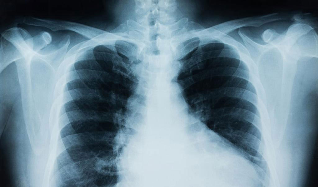
A new method of generating high-quality chest X-ray images can be used to diagnose COVID-19 more accurately than current methods.
The team of researchers at the University of Maryland, Baltimore County (UMBC; Baltimore, MD, USA) has published its findings in the proceedings of the IEEE Big Data 2020 Conference. The need for rapid and accurate COVID-19 testing is high, including testing that can determine if COVID-19 is impacting a patient's respiratory system. Many clinicians use X-ray technology to classify images of possible cases of COVID-19, but the limited data available makes it more challenging to classify those images accurately.
The UMBC researchers developed their tool as an extension of generative adversarial networks (GANs) - machine learning frameworks that can quickly generate new data based on statistics from a training set. The team's more advanced method uses what they call Mean Teacher + Transfer Generative Adversarial Networks (MTT-GAN). The MTT-GANs are superior to GANs because the images they generate are much more similar to authentic images generated by x-ray machines. The MTT-GAN classification system has the potential to help improve the accuracy of COVID-19 classifiers, making it an important diagnostic tool for physicians who are still working to understand the range of ways this complex disease presents in patients.
"The availability of data is one of the most important aspects of machine learning and our research has taken an incremental theoretical step towards generating data using the MTT-GAN," said Sumeet Menon, a Ph.D. student in computer science at UMBC who led the research team. "This paper mainly focuses on generating more COVID-19 X-rays using the MTT-GAN, which could be widely used to train machine learning models and could have many applications, including classification of CT-scans and segmentation."
Related Links:
University of Maryland, Baltimore County





The team of researchers at the University of Maryland, Baltimore County (UMBC; Baltimore, MD, USA) has published its findings in the proceedings of the IEEE Big Data 2020 Conference. The need for rapid and accurate COVID-19 testing is high, including testing that can determine if COVID-19 is impacting a patient's respiratory system. Many clinicians use X-ray technology to classify images of possible cases of COVID-19, but the limited data available makes it more challenging to classify those images accurately.
The UMBC researchers developed their tool as an extension of generative adversarial networks (GANs) - machine learning frameworks that can quickly generate new data based on statistics from a training set. The team's more advanced method uses what they call Mean Teacher + Transfer Generative Adversarial Networks (MTT-GAN). The MTT-GANs are superior to GANs because the images they generate are much more similar to authentic images generated by x-ray machines. The MTT-GAN classification system has the potential to help improve the accuracy of COVID-19 classifiers, making it an important diagnostic tool for physicians who are still working to understand the range of ways this complex disease presents in patients.
"The availability of data is one of the most important aspects of machine learning and our research has taken an incremental theoretical step towards generating data using the MTT-GAN," said Sumeet Menon, a Ph.D. student in computer science at UMBC who led the research team. "This paper mainly focuses on generating more COVID-19 X-rays using the MTT-GAN, which could be widely used to train machine learning models and could have many applications, including classification of CT-scans and segmentation."
Related Links:
University of Maryland, Baltimore County

Gold Member
SARS‑CoV‑2/Flu A/Flu B/RSV Sample-To-Answer Test
SARS‑CoV‑2/Flu A/Flu B/RSV Cartridge (CE-IVD)
Surgical System
Stealth AXiS
Blood Pressure Monitor
Cuff Blood Pressure Monitor
Latest COVID-19 News
- Low-Cost System Detects SARS-CoV-2 Virus in Hospital Air Using High-Tech Bubbles
- World's First Inhalable COVID-19 Vaccine Approved in China
- COVID-19 Vaccine Patch Fights SARS-CoV-2 Variants Better than Needles
- Blood Viscosity Testing Can Predict Risk of Death in Hospitalized COVID-19 Patients
- ‘Covid Computer’ Uses AI to Detect COVID-19 from Chest CT Scans
- MRI Lung-Imaging Technique Shows Cause of Long-COVID Symptoms
- Chest CT Scans of COVID-19 Patients Could Help Distinguish Between SARS-CoV-2 Variants
- Specialized MRI Detects Lung Abnormalities in Non-Hospitalized Long COVID Patients
- AI Algorithm Identifies Hospitalized Patients at Highest Risk of Dying From COVID-19
- Sweat Sensor Detects Key Biomarkers That Provide Early Warning of COVID-19 and Flu
- Study Assesses Impact of COVID-19 on Ventilation/Perfusion Scintigraphy
- CT Imaging Study Finds Vaccination Reduces Risk of COVID-19 Associated Pulmonary Embolism
- Third Day in Hospital a ‘Tipping Point’ in Severity of COVID-19 Pneumonia
- Longer Interval Between COVID-19 Vaccines Generates Up to Nine Times as Many Antibodies
- AI Model for Monitoring COVID-19 Predicts Mortality Within First 30 Days of Admission
- AI Predicts COVID Prognosis at Near-Expert Level Based Off CT Scans
Channels
Artificial Intelligence
view channel
Explainable AI Dermoscopy Tool Supports Risk Assessment for Skin Cancer
Skin lesions can be difficult to triage when benign and malignant findings share visual features, leaving clinicians uncertain about whether to recommend biopsy or continued monitoring. These decisions... Read more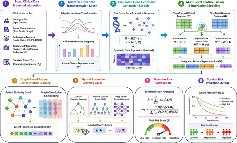
AI Model Improves Breast Cancer Survival Prediction
Predicting survival in breast cancer can be challenging because outcomes are influenced by a combination of clinical, biological, and genetic factors. Traditional survival models can estimate risk but... Read more
Critical Care
view channel
U.S. Launch Expands Kidney Replacement Therapy Options With Integrated Anticoagulation
Critically ill patients with acute kidney injury often require continuous kidney replacement therapy in intensive care units. During these extracorporeal treatments, careful anticoagulation management... Read more
Noninvasive Brain-Computer Interface Enables Responses in Some Unresponsive ICU Patients
Cognitive motor dissociation occurs when patients retain signs of consciousness but cannot move or speak to communicate. In the ICU, this hidden consciousness can be difficult to distinguish from coma... Read more

Surgical Techniques
view channel
Miniature Image Sensor Enables Digital Imaging in Single-Use Endoscopes
Disposable single-use endoscopes are increasingly used to help reduce infection risk and the burden of post-procedure reprocessing and equipment management. As insertion sections become thinner, endoscopic... Read more
Half-Matched Family Donors Could Expand Transplant Access for Severe Aplastic Anemia
Severe aplastic anemia is a rare, life-threatening disorder in which the bone marrow fails to produce enough healthy blood cells, leaving children and young adults vulnerable to anemia, infection, and bleeding.... Read more
Patient Care
view channel
Expert Review Outlines Diagnosis and Treatment of Recurrent Catheter Infections
Patients who rely on long-term intravenous catheters for chemotherapy, hemodialysis, intravenous nutrition, and other essential therapies remain at risk of catheter-related bloodstream infections.... Read more
Total-Body PET/CT Scanner Enables One-Minute Diagnostic-Quality Cancer Imaging
Positron emission tomography/computed tomography (PET/CT) combines radiotracer-based functional imaging with anatomical detail to diagnose, stage, and monitor cancer. Conventional scanners image the body... Read more

Medical Imaging
view channel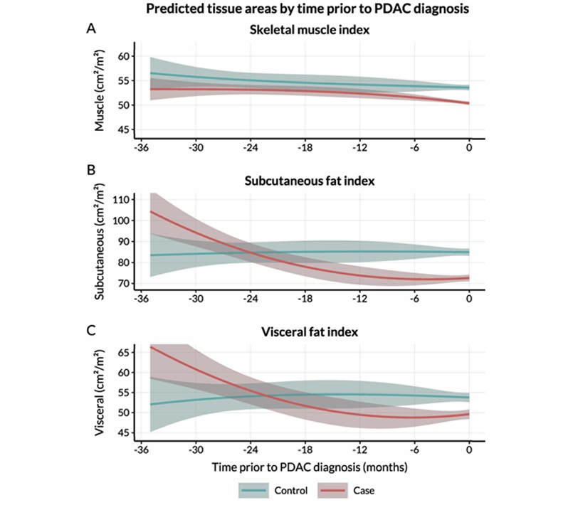
Routine CT Body Composition Tool Helps Identify Early Pancreatic Cancer Risk
Pancreatic ductal adenocarcinoma (PDAC), the most common type of pancreatic cancer, often goes undetected until it has spread. Surveillance programs primarily target people with inherited genetic risk... Read more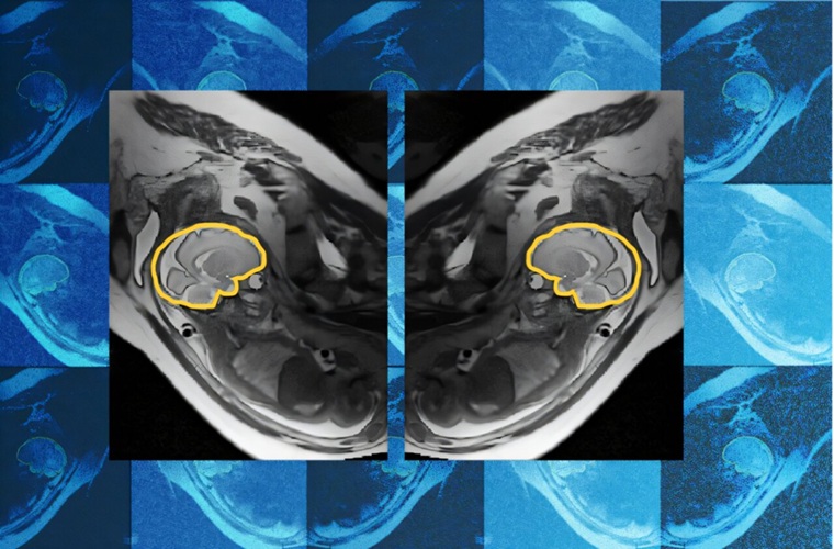
AI Tool Improves Fetal MRI for Brain Abnormality Diagnosis
Fetal brain abnormalities can be difficult to diagnose before birth when fetal motion reduces imaging quality. Routine ultrasound may flag a possible abnormality, prompting follow-up fetal MRI, but head... Read more
AI Model Detects Hidden Disease Signals in Routine CT Scans
Computed tomography (CT) scans are often performed to answer a specific diagnostic question, which can leave other potentially important findings outside the immediate clinical focus. As a result, opportunities... Read more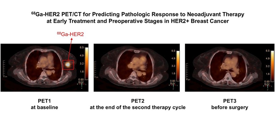
Targeted PET/CT Predicts Early Treatment Response in HER2-Positive Breast Cancer
HER2-positive breast cancer is an aggressive subtype that accounts for a substantial proportion of breast cancer cases. Patients often receive neoadjuvant therapy before surgery to shrink tumors, but assessing... Read moreHealth IT
view channel
Clinical Decision Support Tool Automates CKD Risk Assessment in Primary Care
Chronic kidney disease (CKD) requires timely diagnosis, risk assessment, and coordinated treatment in primary care. Delayed recognition can affect decisions about testing, blood pressure control, cardiovascular... Read more
EHR Alerts Support Dosing, Sedation, and Shock Recognition in Cardiac Care
Electronic health record alerts can add to clinician workload when they are poorly targeted. In critical cardiac care, however, delayed decisions can affect treatment timing, sedation exposure, and escalation... Read more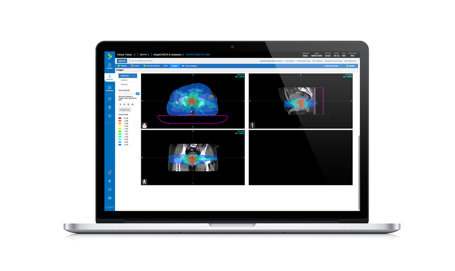
Point of Care
view channel
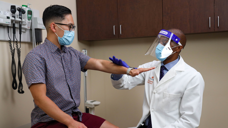
Continuous Glucose Monitoring Identifies Cardiometabolic Risk in Adults Without Diabetes
Dysglycemia—abnormal blood glucose regulation—can fluctuate throughout the day and often escape conventional screening. Clinicians typically rely on fasting plasma glucose and hemoglobin A1c, which offer... Read more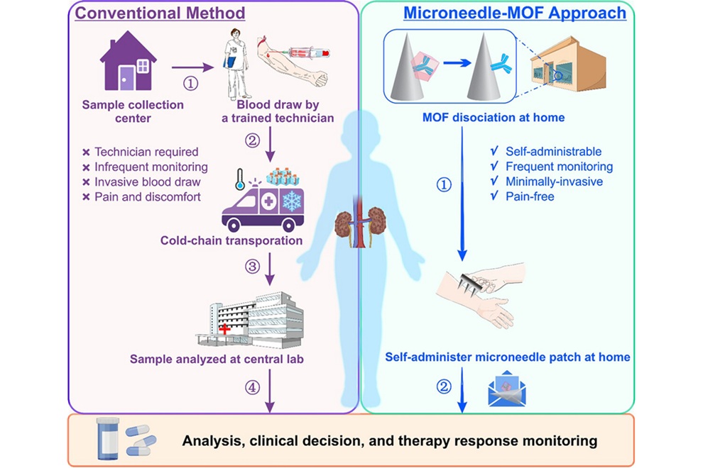
Microneedle Patch Enables At-Home Monitoring of Acute Kidney Injury
Kidney disease is often silent in its early stages, making timely detection difficult outside clinical settings. Early biomarkers such as neutrophil gelatinase–associated lipocalin can require blood draws... Read more
Business
view channel
Steris to Expand Surgical Safety Portfolio With $2.1 Billion Xodus Medical Deal
Steris has signed a definitive agreement to acquire Xodus Medical, a specialty surgical products business based in Pennsylvania, for $2.1 billion at closing. The acquisition will expand Steris’s healthcare... Read more
Stryker Enters Soft-Tissue Robotics Through Strategic Alliance With CMR Surgical
Stryker (Portage, MI, USA) and CMR Surgical (Cambridge, UK) have announced a strategic alliance centered on the Versius Surgical Robotic portfolio. The agreement includes an approximately USD 400 million... Read more
Terumo Neuro Acquisition Advances Liquid Embolic Technology for Chronic Subdural Hematoma
Terumo Neuro (Aliso Viejo, CA, USA) signed an agreement to acquire Arsenal Medical Inc. (Waltham, MA, USA), a privately held, clinical-stage biomaterials company. The proposed acquisition would add NeoCast,... Read more