


Serious COVID-19 Patients Requiring Brain Imaging Face Higher Risk of Death, Finds Study
|
By HospiMedica International staff writers Posted on 24 Dec 2020 |


|

Illustration
Hospitalized COVID-19 patients with neurological problems serious enough to warrant brain imaging have a higher risk of dying, according to a new study.
These findings by researchers at Montefiore Health System (New York City, NY, USA) and Albert Einstein College of Medicine (New York City, NY, USA) have the potential to identify and focus treatment efforts on individuals most at risk and could decrease COVID-19 deaths.
The study looked at data from 4,711 COVID-19 patients who were admitted to Montefiore during the six-week period between March 1, 2020 and April 16, 2020. Of those patients, 581 (12%) had neurological problems serious enough to warrant brain imaging. These individuals were compared with 1,743 non-neurological COVID-19 patients of similar age and disease severity who were admitted during the same period. Among people who underwent brain imaging, 55 were diagnosed with stroke and 258 people exhibited confusion or altered thinking ability. Individuals with stroke were twice as likely to die (49% mortality) compared with their matched controls (24% mortality) - a statistically significant difference. People with confusion had a 40% mortality rate compared with 33% for their matched controls - also statistically significant. More than half the stroke patients in the study did not have hypertension or other underlying risk factors for stroke.
“This study is the first to show that the presence of neurological symptoms, particularly stroke and confused or altered thinking, may indicate a more serious course of illness, even when pulmonary problems aren’t severe,” said David Altschul, M.D., chief of the division of neurovascular surgery at Einstein and Montefiore, and associate professor in the Leo M. Davidoff Department of Neurological Surgery and of radiology at Einstein. “Hospitals can use this knowledge to prioritize treatment and, hopefully, save more lives during this pandemic.”
Related Links:
Montefiore Health System
Albert Einstein College of Medicine





These findings by researchers at Montefiore Health System (New York City, NY, USA) and Albert Einstein College of Medicine (New York City, NY, USA) have the potential to identify and focus treatment efforts on individuals most at risk and could decrease COVID-19 deaths.
The study looked at data from 4,711 COVID-19 patients who were admitted to Montefiore during the six-week period between March 1, 2020 and April 16, 2020. Of those patients, 581 (12%) had neurological problems serious enough to warrant brain imaging. These individuals were compared with 1,743 non-neurological COVID-19 patients of similar age and disease severity who were admitted during the same period. Among people who underwent brain imaging, 55 were diagnosed with stroke and 258 people exhibited confusion or altered thinking ability. Individuals with stroke were twice as likely to die (49% mortality) compared with their matched controls (24% mortality) - a statistically significant difference. People with confusion had a 40% mortality rate compared with 33% for their matched controls - also statistically significant. More than half the stroke patients in the study did not have hypertension or other underlying risk factors for stroke.
“This study is the first to show that the presence of neurological symptoms, particularly stroke and confused or altered thinking, may indicate a more serious course of illness, even when pulmonary problems aren’t severe,” said David Altschul, M.D., chief of the division of neurovascular surgery at Einstein and Montefiore, and associate professor in the Leo M. Davidoff Department of Neurological Surgery and of radiology at Einstein. “Hospitals can use this knowledge to prioritize treatment and, hopefully, save more lives during this pandemic.”
Related Links:
Montefiore Health System
Albert Einstein College of Medicine

Gold Member
Blood Gas Analyzer
i-Check200
Digital Radiography System (Ceiling Free)
Digix CF Series
Wound Irrigation Solution
Prontosan®
Latest COVID-19 News
- Low-Cost System Detects SARS-CoV-2 Virus in Hospital Air Using High-Tech Bubbles
- World's First Inhalable COVID-19 Vaccine Approved in China
- COVID-19 Vaccine Patch Fights SARS-CoV-2 Variants Better than Needles
- Blood Viscosity Testing Can Predict Risk of Death in Hospitalized COVID-19 Patients
- ‘Covid Computer’ Uses AI to Detect COVID-19 from Chest CT Scans
- MRI Lung-Imaging Technique Shows Cause of Long-COVID Symptoms
- Chest CT Scans of COVID-19 Patients Could Help Distinguish Between SARS-CoV-2 Variants
- Specialized MRI Detects Lung Abnormalities in Non-Hospitalized Long COVID Patients
- AI Algorithm Identifies Hospitalized Patients at Highest Risk of Dying From COVID-19
- Sweat Sensor Detects Key Biomarkers That Provide Early Warning of COVID-19 and Flu
- Study Assesses Impact of COVID-19 on Ventilation/Perfusion Scintigraphy
- CT Imaging Study Finds Vaccination Reduces Risk of COVID-19 Associated Pulmonary Embolism
- Third Day in Hospital a ‘Tipping Point’ in Severity of COVID-19 Pneumonia
- Longer Interval Between COVID-19 Vaccines Generates Up to Nine Times as Many Antibodies
- AI Model for Monitoring COVID-19 Predicts Mortality Within First 30 Days of Admission
- AI Predicts COVID Prognosis at Near-Expert Level Based Off CT Scans
Channels
Artificial Intelligence
view channel
AI Improves Non-Contrast CT Interpretation for Time-Sensitive Stroke Assessment
Acute ischemic stroke occurs when a blood vessel in the brain becomes blocked, requiring rapid diagnosis to enable timely reperfusion therapy. Emergency departments often use computed tomography angiography... Read more
AI-Powered ECG Model Gains FDA Authorization for Emergency Cardiac Assessment
Powerful Medical’s PMcardio Queen of Hearts (ACS AI ECG Model) has received U.S. Food and Drug Administration (FDA) De Novo authorization, announced on September 15, 2026. The model is designed to identify... Read more
Critical Care
view channel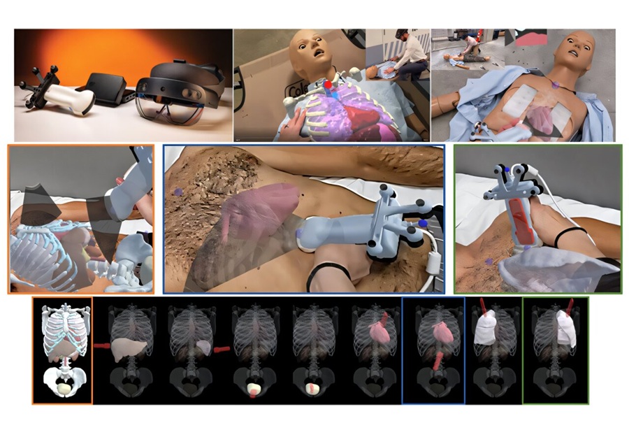
Anatomical Guidance System Improves Trauma Ultrasound Acquisition by Novice Users
Focused assessment with sonography for trauma (FAST) helps clinicians detect free fluid and signs of internal bleeding in injured patients, but obtaining reliable images can be difficult for inexperienced operators.... Read more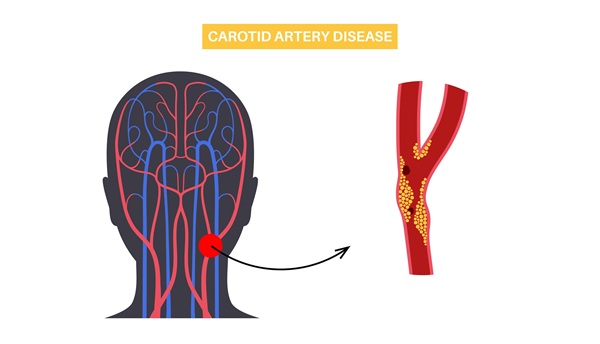
AI-Enhanced Handheld Ultrasound Improves Carotid Plaque Detection
Handheld ultrasound can miss small or faint carotid plaque, creating uncertainty in community screening. This gap makes it harder for frontline clinicians to decide who needs confirmatory imaging, closer... Read more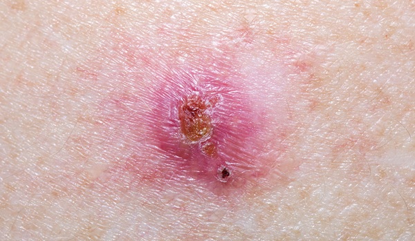
Noninvasive Imaging Approach Aims to Detect Basal Cell Carcinoma Before It Becomes Visible
Basal cell carcinoma is the most common form of skin cancer and can damage nearby structures such as the nose or eyes when it develops on the face. Diagnosis typically depends on visible skin changes,... Read more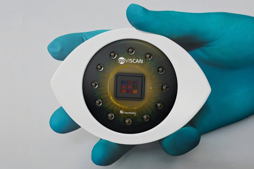
Surgical Techniques
view channel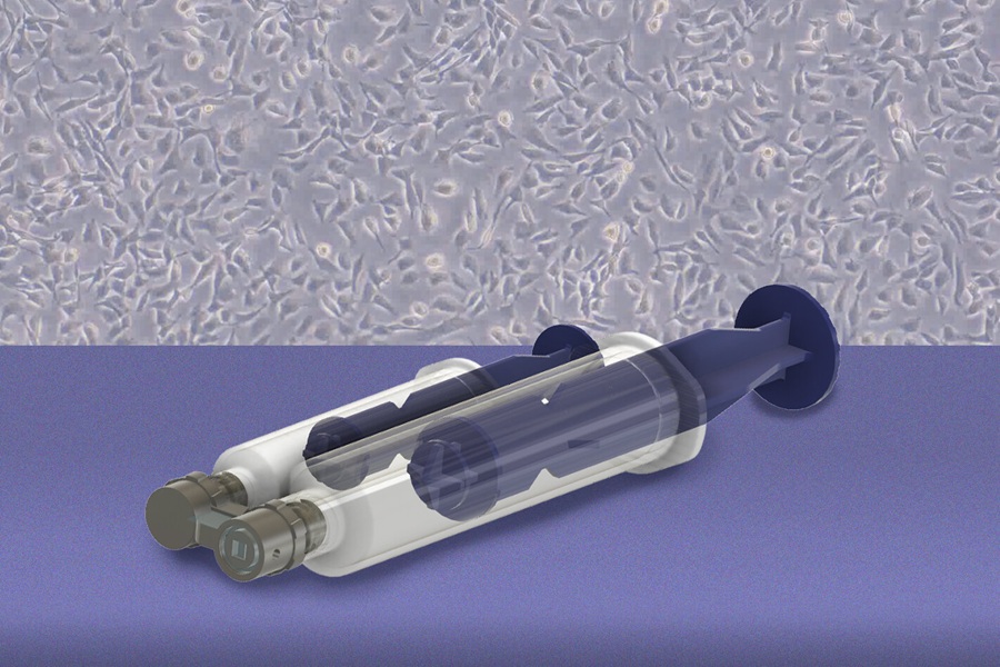
Handheld Device Improves Targeted Cell Collection for Early Ovarian Cancer Assessment
High-grade serous ovarian cancer is the most common form of ovarian cancer, and many cases are thought to originate in the fallopian tubes. Because precursor lesions can be microscopic, they are difficult... Read more
Light-Activated Nanoparticles May Offer New Approach to Retinal Prostheses
Retinitis pigmentosa is a degenerative retinal disorder in which photoreceptors progressively die, reducing visual signals to the brain while leaving surviving inner retinal circuits underused.... Read more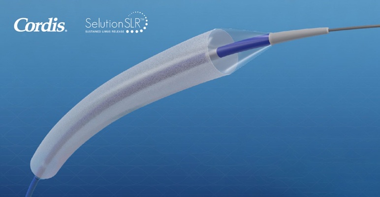
FDA Approves First Sirolimus Drug-Eluting Balloon for Coronary In-Stent Restenosis
Coronary in-stent restenosis (ISR) accounts for about 10% of the nearly one million percutaneous coronary interventions performed annually in the United States. Treating recurrent narrowing without another... Read more
Patient Care
view channel
EHR Model Flags High-Risk Periods in Patients with Metastatic Breast Cancer
Determining when patients with metastatic breast cancer are nearing the end of life remains difficult, often leading to intensive interventions with limited benefit. Prognostic uncertainty can delay goals-of-care... Read more
Virtual Reality Helps Claustrophobic Patients Complete Cardiac PET/CT Scans
Anxiety and claustrophobia—intense fear of confined spaces—often prevent patients from completing cardiac positron emission tomography/computed tomography (PET/CT). Aborted scans delay diagnosis, consume... Read more

Medical Imaging
view channel
New MRI Approach Captures Rapidly Decaying Tissue Signals
MRI measures signals generated as excited proton spins return toward equilibrium. However, some tissue signals decay so rapidly that conventional systems can miss them during the brief interval after excitation,... Read more
AI Tool Integrates MRI and Ultrasound to Detect Endometriosis
Endometriosis is a chronic condition in which tissue similar to the uterine lining grows outside the uterus, causing pain, heavy bleeding, and infertility. Diagnosis often depends on surgical visualization,... Read more

Health IT
view channel
New Tablet Platform Extends Stroke Neurorehabilitation Beyond the Clinic
Continuity and intensity of neurorehabilitation after stroke are critical for functional recovery, but access to ongoing therapy at home often remains limited. Hospitals and outpatient programs therefore... Read more
Smartphone App Improves Blood Pressure Control in Hypertension
Hypertension, or high blood pressure, is a major modifiable driver of cardiovascular, renal, and cerebrovascular morbidity. Despite effective therapies, many adults fail to achieve control, leading to... Read more

Point of Care
view channel
Continuous Glucose Monitoring Identifies Cardiometabolic Risk in Adults Without Diabetes
Dysglycemia—abnormal blood glucose regulation—can fluctuate throughout the day and often escape conventional screening. Clinicians typically rely on fasting plasma glucose and hemoglobin A1c, which offer... Read more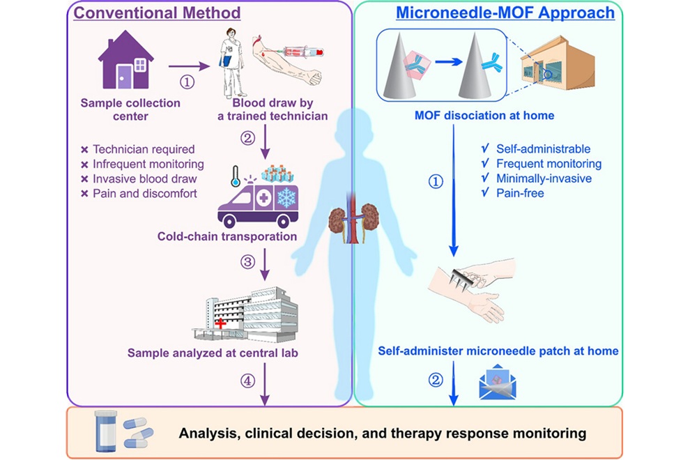
Microneedle Patch Enables At-Home Monitoring of Acute Kidney Injury
Kidney disease is often silent in its early stages, making timely detection difficult outside clinical settings. Early biomarkers such as neutrophil gelatinase–associated lipocalin can require blood draws... Read more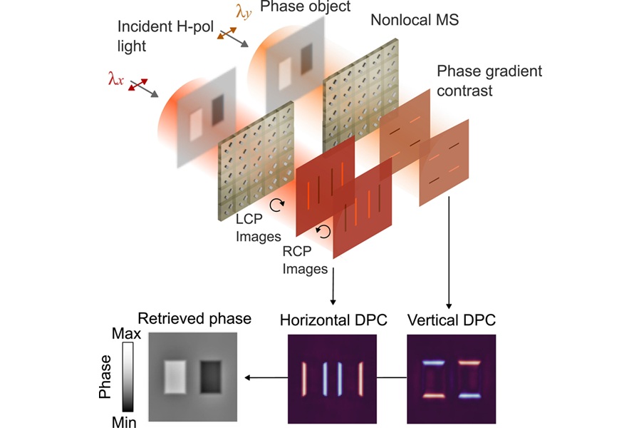
Business
view channel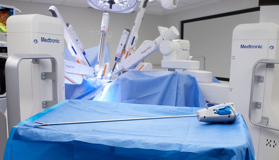
Medtronic Receives FDA Clearance for Vessel-Sealing Instrument for Robotic Surgery
As robotic-assisted surgery expands across U.S. hospitals, teams increasingly seek energy instruments with the familiarity and performance of tools used in open and laparoscopic procedures.... Read more
Robotic Single-Port System Gains CE Mark for Transvaginal Gynecologic Procedures
Intuitive (Sunnyvale, CA, USA) announced that it has received CE mark approval for use of the da Vinci SP Single Port surgical system in transvaginal gynecologic procedures, marking the first such indication... Read more