Lung CT Scans of COVID-19 Patients Can Predict Neurological Problems Revealed Later by Brain MRIs
|
By HospiMedica International staff writers Posted on 15 Mar 2021 |

Image: Achala Vagal, MD, author on the study, reviews an MRI scan (Photo courtesy of Colleen Kelley)
For the first time, a multicenter study has found a visual correlation between the severity of the disease in the lungs using CT scans and the severity of effects on patient’s brains, using MRI scans.
The results of the study led by University of Cincinnati (Cincinnati, OH, USA) show that by looking at lung CT scans of patients diagnosed with COVID-19, physicians may be able to predict just how badly they can experience other neurological problems that could show up on brain MRIs, helping improve patient outcomes and identify symptoms for earlier treatment. CT imaging can detect illness in the lungs better than an MRI, another medical imaging technique. However, MRI can detect many problems in the brain, particularly in COVID-19 patients, that cannot be detected on CT images.
In the study, researchers reviewed electronic medical records and images of hospitalized COVID-19 patients. Patients who were diagnosed with COVID-19, experienced neurological issues and who had both lung and brain images available were included. Out of 135 COVID-19 patients with abnormal CT lung scans and neurological symptoms, 49, or 36%, were also found to develop abnormal brain scans and were more likely to experience stroke symptoms.
The researchers believe that the study will help physicians classify patients, based on the severity of disease found on their CT scans, into groups more likely to develop brain imaging abnormalities. Additionally, the correlation could be important for implementing therapies, particularly in stroke prevention, to improve outcomes in patients with COVID-19.
“These results are important because they further show that severe lung disease from COVID-19 could mean serious brain complications, and we have the imaging to help prove it,” said Abdelkader Mahammedi, MD, assistant professor of radiology, UC Health radiologist and member of the UC Gardner Neuroscience Institute. “Future larger studies are needed to help us understand the tie better, but for now, we hope these results can be used to help predict care and ensure that patients have the best outcomes.”
Related Links:
University of Cincinnati
Latest COVID-19 News
- Low-Cost System Detects SARS-CoV-2 Virus in Hospital Air Using High-Tech Bubbles
- World's First Inhalable COVID-19 Vaccine Approved in China
- COVID-19 Vaccine Patch Fights SARS-CoV-2 Variants Better than Needles
- Blood Viscosity Testing Can Predict Risk of Death in Hospitalized COVID-19 Patients
- ‘Covid Computer’ Uses AI to Detect COVID-19 from Chest CT Scans
- MRI Lung-Imaging Technique Shows Cause of Long-COVID Symptoms
- Chest CT Scans of COVID-19 Patients Could Help Distinguish Between SARS-CoV-2 Variants
- Specialized MRI Detects Lung Abnormalities in Non-Hospitalized Long COVID Patients
- AI Algorithm Identifies Hospitalized Patients at Highest Risk of Dying From COVID-19
- Sweat Sensor Detects Key Biomarkers That Provide Early Warning of COVID-19 and Flu
- Study Assesses Impact of COVID-19 on Ventilation/Perfusion Scintigraphy
- CT Imaging Study Finds Vaccination Reduces Risk of COVID-19 Associated Pulmonary Embolism
- Third Day in Hospital a ‘Tipping Point’ in Severity of COVID-19 Pneumonia
- Longer Interval Between COVID-19 Vaccines Generates Up to Nine Times as Many Antibodies
- AI Model for Monitoring COVID-19 Predicts Mortality Within First 30 Days of Admission
- AI Predicts COVID Prognosis at Near-Expert Level Based Off CT Scans
Channels
Critical Care
view channel
AI Tool Improves Speed and Accuracy of Cervical Cancer Treatment Planning
Cervical cancer affects around 600,000 women globally each year and causes approximately 340,000 deaths. Brachytherapy is among the most effective treatments for this cancer, but its use is often limited... Read more
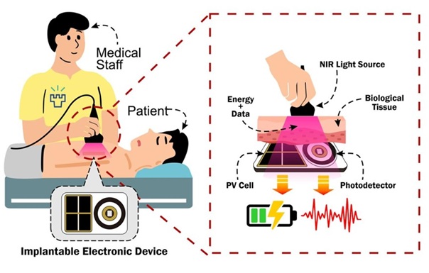
Ultrasonic Sensor Enables Cuffless and Non-Invasive Blood Pressure Measurement
Accurate blood pressure monitoring is essential for managing cardiovascular disease, yet conventional cuff-based systems are bulky, intermittent, and uncomfortable for long-term use. Optical cuffless methods... Read moreSurgical Techniques
view channel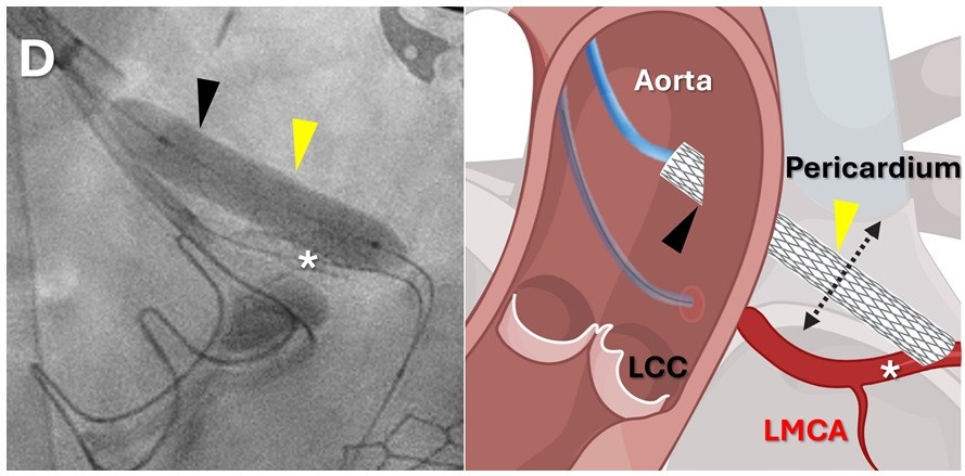
Minimally Invasive Coronary Artery Bypass Method Offers Safer Alternative to Open-Heart Surgery
Coronary artery obstruction is a rare but often fatal complication of heart-valve replacement, particularly in patients with complex anatomy or prior cardiac interventions. In such cases, traditional open-heart... Read more
Injectable Breast ‘Implant’ Offers Alternative to Traditional Surgeries
Breast cancer surgery can require the removal of part or all of the breast, leaving patients with difficult decisions about reconstruction. Current reconstructive options often rely on prosthetic implants... Read morePatient Care
view channel
Revolutionary Automatic IV-Line Flushing Device to Enhance Infusion Care
More than 80% of in-hospital patients receive intravenous (IV) therapy. Every dose of IV medicine delivered in a small volume (<250 mL) infusion bag should be followed by subsequent flushing to ensure... Read more
VR Training Tool Combats Contamination of Portable Medical Equipment
Healthcare-associated infections (HAIs) impact one in every 31 patients, cause nearly 100,000 deaths each year, and cost USD 28.4 billion in direct medical expenses. Notably, up to 75% of these infections... Read more
Portable Biosensor Platform to Reduce Hospital-Acquired Infections
Approximately 4 million patients in the European Union acquire healthcare-associated infections (HAIs) or nosocomial infections each year, with around 37,000 deaths directly resulting from these infections,... Read moreFirst-Of-Its-Kind Portable Germicidal Light Technology Disinfects High-Touch Clinical Surfaces in Seconds
Reducing healthcare-acquired infections (HAIs) remains a pressing issue within global healthcare systems. In the United States alone, 1.7 million patients contract HAIs annually, leading to approximately... Read moreHealth IT
view channel
EMR-Based Tool Predicts Graft Failure After Kidney Transplant
Kidney transplantation offers patients with end-stage kidney disease longer survival and better quality of life than dialysis, yet graft failure remains a major challenge. Although a successful transplant... Read more
Printable Molecule-Selective Nanoparticles Enable Mass Production of Wearable Biosensors
The future of medicine is likely to focus on the personalization of healthcare—understanding exactly what an individual requires and delivering the appropriate combination of nutrients, metabolites, and... Read moreBusiness
view channel
Philips and Masimo Partner to Advance Patient Monitoring Measurement Technologies
Royal Philips (Amsterdam, Netherlands) and Masimo (Irvine, California, USA) have renewed their multi-year strategic collaboration, combining Philips’ expertise in patient monitoring with Masimo’s noninvasive... Read more
B. Braun Acquires Digital Microsurgery Company True Digital Surgery
The high-end microsurgery market in neurosurgery, spine, and ENT is undergoing a significant transformation. Traditional analog microscopes are giving way to digital exoscopes, which provide improved visualization,... Read more
CMEF 2025 to Promote Holistic and High-Quality Development of Medical and Health Industry
The 92nd China International Medical Equipment Fair (CMEF 2025) Autumn Exhibition is scheduled to be held from September 26 to 29 at the China Import and Export Fair Complex (Canton Fair Complex) in Guangzhou.... Read more


