Highly Advanced AI Model Precisely Detects COVID-19 by Analyzing Lung Images
|
By HospiMedica International staff writers Posted on 13 Aug 2021 |
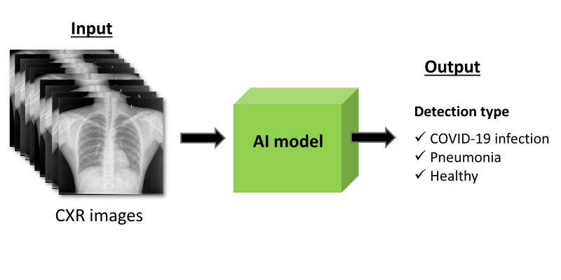
Illustration
Researchers have designed and validated an image-based detection of COVID-19 with the aid of artificial intelligence (AI) models by using a model to automatically collect imaging data from the lung lobes. This data was then analyzed to yield features as potential diagnostic biomarkers for COVID-19.
These diagnostic biomarkers using the AI model were subsequently used to differentiate COVID-19 patients from both pneumonia and healthy patients. The entire model was developed by researchers from the Terasaki Institute for Biomedical Innovation (TIBI; Los Angeles, CA, USA) with a cohort of 704 chest X-rays and then independently validated with 1,597 cases from multiple sources comprised of healthy, pneumonia, and COVID-19 patients. The results showed excellent performance by the model in classifying diagnoses of the various patients.
Medical imaging has long been a vital tool for the diagnosis and prognostic assessments of many diseases. In recent years, the use of AI models has been used in conjunction with this imaging to augment their diagnostic capabilities. By using these models, some features can be extracted from images that may reveal disease characteristics not identified by the naked eye. The power to process data in this intelligent manner can have a big impact on the medical field, especially with the current growth in imaging features and the need for high precision in medical decisions.
There is a huge demand for rapid and accurate detection of COVID-19 infection. The primary detection method has been using reverse transcription-polymerase chain reaction (RT-PCR) on samples collected from nasal or throat swabs. However, this method is subject to inaccuracies due to sampling errors, low viral load, and the method’s sensitivity limitations. This is an especially significant issue for patients who are in the early stages of infection. An additional diagnostic tool for COVID-19 can come from images of lungs. For diagnosing lung diseases, chest X-rays or CT scans are the primary resources, and they can be used to distinguish COVID-19 from other types of lung injuries, as well as to assess the severity of lung involvement in COVID-19. These types of images can enhance the diagnostic capabilities for COVID-19 patients, especially if they are coupled with AI models. The use of computer modeling with data extracted from medical images shows great promise in enabling precision medicine and can revolutionize medical practice in the clinic. Developing methodologies to capture entire sets of information while suppressing irrelevant features enhances the reliability of AI models. The proposed approach would be a step towards applying them in precision medicine and can provide an efficient, inexpensive, and non-invasive way to strengthen the diagnostic capabilities of imaging.
“This highly advanced artificial intelligence model further helps our ability to precisely detect COVID-19 patients. In addition, such a model can be applied for diagnosis of other diseases using different imaging modalities,” said lead researcher Samad Ahadian, Ph.D.
“Artificial intelligence-driven models with diagnostic and predictive capabilities are a powerful tool that are an important part of our research platforms here at the institute,” said Ali Khademhosseini, Ph.D., Director and CEO of TIBI. “This will carry over into countless applications in the biomedical field and in the clinic.”
Related Links:
Terasaki Institute for Biomedical Innovation
Latest COVID-19 News
- Low-Cost System Detects SARS-CoV-2 Virus in Hospital Air Using High-Tech Bubbles
- World's First Inhalable COVID-19 Vaccine Approved in China
- COVID-19 Vaccine Patch Fights SARS-CoV-2 Variants Better than Needles
- Blood Viscosity Testing Can Predict Risk of Death in Hospitalized COVID-19 Patients
- ‘Covid Computer’ Uses AI to Detect COVID-19 from Chest CT Scans
- MRI Lung-Imaging Technique Shows Cause of Long-COVID Symptoms
- Chest CT Scans of COVID-19 Patients Could Help Distinguish Between SARS-CoV-2 Variants
- Specialized MRI Detects Lung Abnormalities in Non-Hospitalized Long COVID Patients
- AI Algorithm Identifies Hospitalized Patients at Highest Risk of Dying From COVID-19
- Sweat Sensor Detects Key Biomarkers That Provide Early Warning of COVID-19 and Flu
- Study Assesses Impact of COVID-19 on Ventilation/Perfusion Scintigraphy
- CT Imaging Study Finds Vaccination Reduces Risk of COVID-19 Associated Pulmonary Embolism
- Third Day in Hospital a ‘Tipping Point’ in Severity of COVID-19 Pneumonia
- Longer Interval Between COVID-19 Vaccines Generates Up to Nine Times as Many Antibodies
- AI Model for Monitoring COVID-19 Predicts Mortality Within First 30 Days of Admission
- AI Predicts COVID Prognosis at Near-Expert Level Based Off CT Scans
Channels
Critical Care
view channel
Ingestible Capsule Monitors Intestinal Inflammation
Acute mesenteric ischemia—a life-threatening condition caused by blocked blood flow to the intestines—remains difficult to diagnose early because its symptoms often mimic common digestive problems.... Read more
Wireless Implantable Sensor Enables Continuous Endoleak Monitoring
Endovascular aneurysm repair (EVAR) is a life-saving, minimally invasive treatment for abdominal aortic aneurysms—balloon-like bulges in the aorta that can rupture with fatal consequences.... Read more
Wearable Patch for Early Skin Cancer Detection to Reduce Unnecessary Biopsies
Skin cancer remains one of the most dangerous and common cancers worldwide, with early detection crucial for improving survival rates. Traditional diagnostic methods—visual inspections, imaging, and biopsies—can... Read moreSurgical Techniques
view channel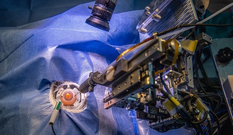
Robotic Assistant Delivers Ultra-Precision Injections with Rapid Setup Times
Age-related macular degeneration (AMD) is a leading cause of blindness worldwide, affecting nearly 200 million people, a figure expected to rise to 280 million by 2040. Current treatment involves doctors... Read more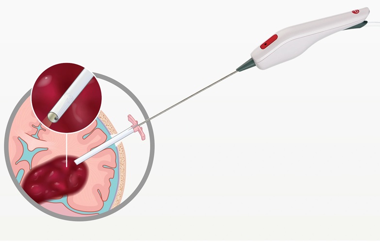
Minimally Invasive Endoscopic Surgery Improves Severe Stroke Outcomes
Intracerebral hemorrhage, a type of stroke caused by bleeding deep within the brain, remains one of the most challenging neurological emergencies to treat. Accounting for about 15% of all strokes, it carries... Read morePatient Care
view channel
Revolutionary Automatic IV-Line Flushing Device to Enhance Infusion Care
More than 80% of in-hospital patients receive intravenous (IV) therapy. Every dose of IV medicine delivered in a small volume (<250 mL) infusion bag should be followed by subsequent flushing to ensure... Read more
VR Training Tool Combats Contamination of Portable Medical Equipment
Healthcare-associated infections (HAIs) impact one in every 31 patients, cause nearly 100,000 deaths each year, and cost USD 28.4 billion in direct medical expenses. Notably, up to 75% of these infections... Read more
Portable Biosensor Platform to Reduce Hospital-Acquired Infections
Approximately 4 million patients in the European Union acquire healthcare-associated infections (HAIs) or nosocomial infections each year, with around 37,000 deaths directly resulting from these infections,... Read moreFirst-Of-Its-Kind Portable Germicidal Light Technology Disinfects High-Touch Clinical Surfaces in Seconds
Reducing healthcare-acquired infections (HAIs) remains a pressing issue within global healthcare systems. In the United States alone, 1.7 million patients contract HAIs annually, leading to approximately... Read moreHealth IT
view channel
Printable Molecule-Selective Nanoparticles Enable Mass Production of Wearable Biosensors
The future of medicine is likely to focus on the personalization of healthcare—understanding exactly what an individual requires and delivering the appropriate combination of nutrients, metabolites, and... Read moreBusiness
view channel
Philips and Masimo Partner to Advance Patient Monitoring Measurement Technologies
Royal Philips (Amsterdam, Netherlands) and Masimo (Irvine, California, USA) have renewed their multi-year strategic collaboration, combining Philips’ expertise in patient monitoring with Masimo’s noninvasive... Read more
B. Braun Acquires Digital Microsurgery Company True Digital Surgery
The high-end microsurgery market in neurosurgery, spine, and ENT is undergoing a significant transformation. Traditional analog microscopes are giving way to digital exoscopes, which provide improved visualization,... Read more
CMEF 2025 to Promote Holistic and High-Quality Development of Medical and Health Industry
The 92nd China International Medical Equipment Fair (CMEF 2025) Autumn Exhibition is scheduled to be held from September 26 to 29 at the China Import and Export Fair Complex (Canton Fair Complex) in Guangzhou.... Read more