



Deep Learning Neural Network Quickly Detects COVID-19 Infections Using X-Ray Images
|
By HospiMedica International staff writers Posted on 24 Nov 2021 |


|

A deep learning neural network can quickly detect COVID-19 infections using X-ray images.
The deep learning neural network named CORONA-Net was developed by scientists at The University of British Columbia (Kelowna, BC Canada) to help doctors who lack access to polymerase chain reaction (PCR) tests and need a way to rapidly screen patients for COVID-19. As COVID-19 continues to make headlines across the globe, people have become used to the idea of rapid testing to determine if they have been infected. The viral test only indicates if a current infection exists, but not if there was previous infection. The alternative antibody test uses a blood sample and can detect if there was a previous infection with the SARS-CoV-2 virus, even if there are no current symptoms. However, the PCR test can be rare in many countries and usually costs several hundred dollars each time. Doctors around the world need a way to rapidly test patients for COVID-19 so that they can begin immediate treatment for patients with the virus
UBC Okanagan researchers, who say rapid tests can be limited and expensive in many countries, are testing another testing method. And they believe, thanks to artificial intelligence, they have found one. The research team has developed CORONA-Net, a deep learning neural network that can quickly detect COVID-19 infections using X-ray images. In many countries, people opt for chest X-ray because of the cost of a PCR test or its unavailability. However, sometimes it is difficult to get the X-ray looked at by a specialist, and accurately detecting the infection can take time. But by using CORONA-NET, the artificial intelligence system can flag suspicious cases to be fast-tracked and looked at quickly.
The developed CORONA-Net architecture substantially increases the sensitivity and positive predictive value (PPV) of predictions, making CORONA-Net a valuable tool when it comes to using chest X-rays to diagnose COVID-19. According to the researchers, the developed CORONA-Net was able to produce results with an accuracy of more than 95% in classifying COVID-19 cases from digital chest X-ray images. The accuracy of detecting COVID-19 by CORONA-Net will continue to increase as the dataset grows. CORONA-Net can automatically improve itself over time and self-learn to be more accurate.
“COVID-19 typically causes pneumonia in human lungs, which can be detected in X-ray images. These datasets of X-rays - of people with pneumonia inflicted by COVID-19, of people with pneumonia inflicted by other diseases, as well as X-rays of healthy people - allow the possibility to create deep learning networks that can differentiate between images of people with COVID-19 and people who do not have the disease,” said graduate student Sherif Elbishlawi, who helped develop CORONA-Net.
“The results on the testing set were obtained and can be seen in 100 per cent sensitivity to the COVID-19 class. There was a 95% sensitivity in the classification of the pneumonia class and a 95 per cent sensitivity in the classification of the normal class,” he added. “These results show that CORONA-Net gives a highly accurate prediction with the most sensitivity to the COVID-19 class.”
Related Links:
The University of British Columbia





Latest COVID-19 News
- Low-Cost System Detects SARS-CoV-2 Virus in Hospital Air Using High-Tech Bubbles
- World's First Inhalable COVID-19 Vaccine Approved in China
- COVID-19 Vaccine Patch Fights SARS-CoV-2 Variants Better than Needles
- Blood Viscosity Testing Can Predict Risk of Death in Hospitalized COVID-19 Patients
- ‘Covid Computer’ Uses AI to Detect COVID-19 from Chest CT Scans
- MRI Lung-Imaging Technique Shows Cause of Long-COVID Symptoms
- Chest CT Scans of COVID-19 Patients Could Help Distinguish Between SARS-CoV-2 Variants
- Specialized MRI Detects Lung Abnormalities in Non-Hospitalized Long COVID Patients
- AI Algorithm Identifies Hospitalized Patients at Highest Risk of Dying From COVID-19
- Sweat Sensor Detects Key Biomarkers That Provide Early Warning of COVID-19 and Flu
- Study Assesses Impact of COVID-19 on Ventilation/Perfusion Scintigraphy
- CT Imaging Study Finds Vaccination Reduces Risk of COVID-19 Associated Pulmonary Embolism
- Third Day in Hospital a ‘Tipping Point’ in Severity of COVID-19 Pneumonia
- Longer Interval Between COVID-19 Vaccines Generates Up to Nine Times as Many Antibodies
- AI Model for Monitoring COVID-19 Predicts Mortality Within First 30 Days of Admission
- AI Predicts COVID Prognosis at Near-Expert Level Based Off CT Scans
Channels
Artificial Intelligence
view channel
AI Chatbot Helps Women Identify Heart Disease Risks and Symptoms
Heart disease is the leading cause of death among women and affects more than 60 million women in the U.S. Despite public health campaigns, awareness of its causes, risk factors, and symptoms has declined,... Read more
AI Tool Prioritizes Peer Referrals to Expand HIV Testing Access
Human immunodeficiency virus (HIV) remains a major public health challenge when people at risk lack timely testing and care. In South Africa, men and adolescents are among those less likely to seek testing... Read more
Critical Care
view channel
Circuit-Free Smart Patch Uses Smartphone Camera to Monitor Pulse and Vessel Stiffness
Routine cardiovascular monitoring often relies on electronic sensors, cables, or specialized equipment, which can limit its use beyond hospitals, ambulances, general practice clinics, and emergency rooms.... Read more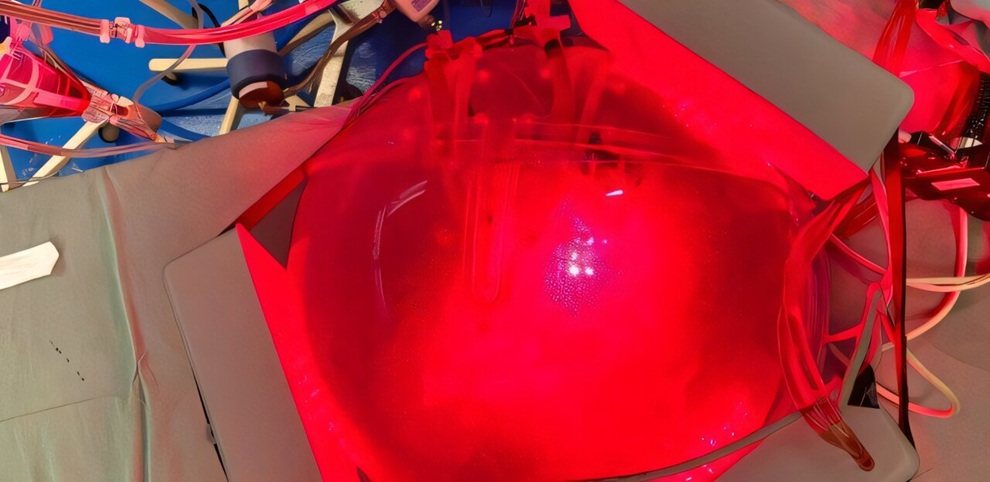
Chest Digital Twin Guides Light Therapy Dosing for Pneumonia
Pneumonia is a lung infection that can impair gas exchange and progress to severe respiratory compromise. Light-based therapies may help treat infection or inflammation, but externally applied light must... Read more
Surgical Techniques
view channel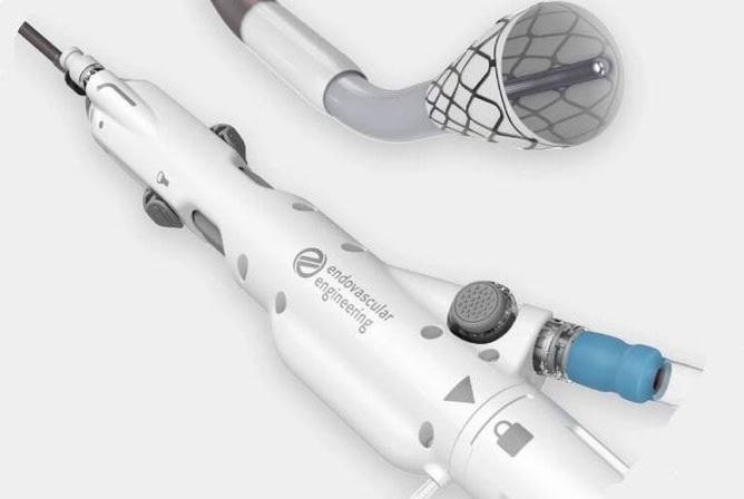
FDA-Cleared Thrombectomy System Removes Pulmonary Embolism Clots
Pulmonary embolism occurs when blood clots travel to the lungs and block blood flow, potentially requiring urgent endovascular intervention to prevent death. Catheter-based clot removal can be challenging,... Read more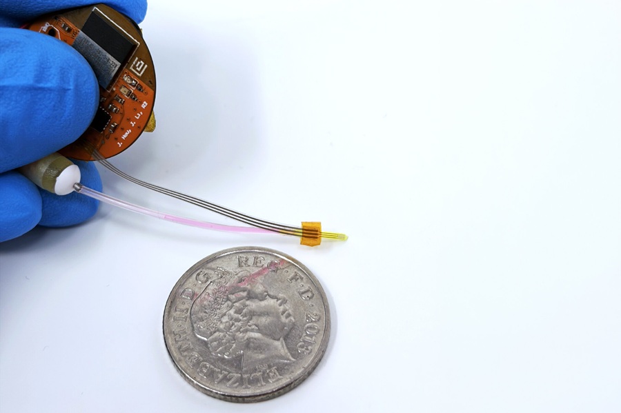
Implantable Ultrasound Device Enhances Local Drug Delivery in Glioblastoma
Glioblastoma is an aggressive brain tumor that frequently recurs after surgery. Although surgeons can remove visible tumor tissue, residual cancer cells may remain in surrounding brain tissue and contribute... Read more
Miniature Image Sensor Enables Digital Imaging in Single-Use Endoscopes
Disposable single-use endoscopes are increasingly used to help reduce infection risk and the burden of post-procedure reprocessing and equipment management. As insertion sections become thinner, endoscopic... Read more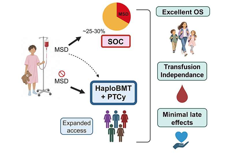
Half-Matched Family Donors Could Expand Transplant Access for Severe Aplastic Anemia
Severe aplastic anemia is a rare, life-threatening disorder in which the bone marrow fails to produce enough healthy blood cells, leaving children and young adults vulnerable to anemia, infection, and bleeding.... Read morePatient Care
view channel
Expert Review Outlines Diagnosis and Treatment of Recurrent Catheter Infections
Patients who rely on long-term intravenous catheters for chemotherapy, hemodialysis, intravenous nutrition, and other essential therapies remain at risk of catheter-related bloodstream infections.... Read more
Total-Body PET/CT Scanner Enables One-Minute Diagnostic-Quality Cancer Imaging
Positron emission tomography/computed tomography (PET/CT) combines radiotracer-based functional imaging with anatomical detail to diagnose, stage, and monitor cancer. Conventional scanners image the body... Read more
Medical Imaging
view channel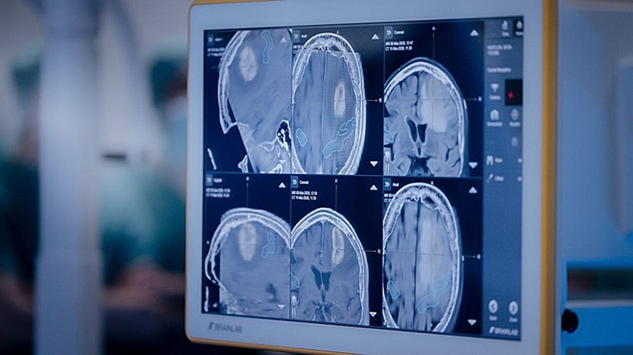
Privacy-Preserving AI Tool Synthesizes CT for Radiation Therapy Planning
Radiation therapy planning depends on accurate medical imaging to calculate dose, visualize organs, and position patients during treatment. Hospitals need large imaging datasets to train robust artificial... Read more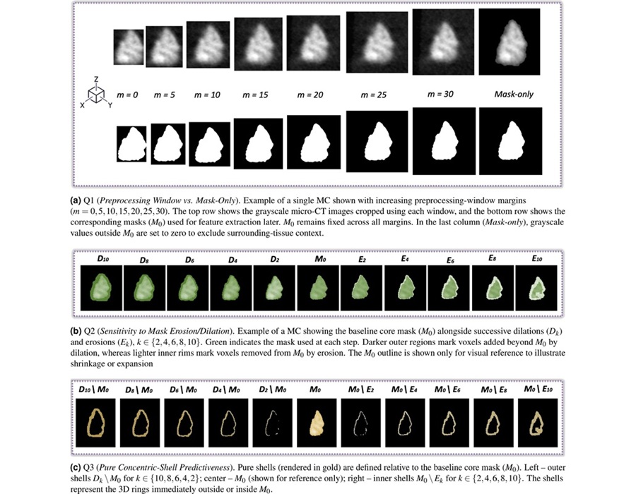
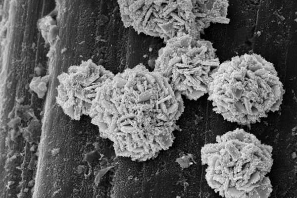
Tissue Around Breast Microcalcifications Emerges as a Key Source of Diagnostic Information
Breast microcalcifications are small calcium deposits that are often detected on mammography and digital breast tomosynthesis. Clinicians use their morphology and distribution to assess suspicious breast... Read more

Health IT
view channel
Clinical Decision Support Tool Automates CKD Risk Assessment in Primary Care
Chronic kidney disease (CKD) requires timely diagnosis, risk assessment, and coordinated treatment in primary care. Delayed recognition can affect decisions about testing, blood pressure control, cardiovascular... Read more
EHR Alerts Support Dosing, Sedation, and Shock Recognition in Cardiac Care
Electronic health record alerts can add to clinician workload when they are poorly targeted. In critical cardiac care, however, delayed decisions can affect treatment timing, sedation exposure, and escalation... Read more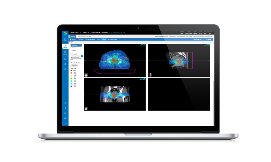
Point of Care
view channel
Continuous Glucose Monitoring Identifies Cardiometabolic Risk in Adults Without Diabetes
Dysglycemia—abnormal blood glucose regulation—can fluctuate throughout the day and often escape conventional screening. Clinicians typically rely on fasting plasma glucose and hemoglobin A1c, which offer... Read more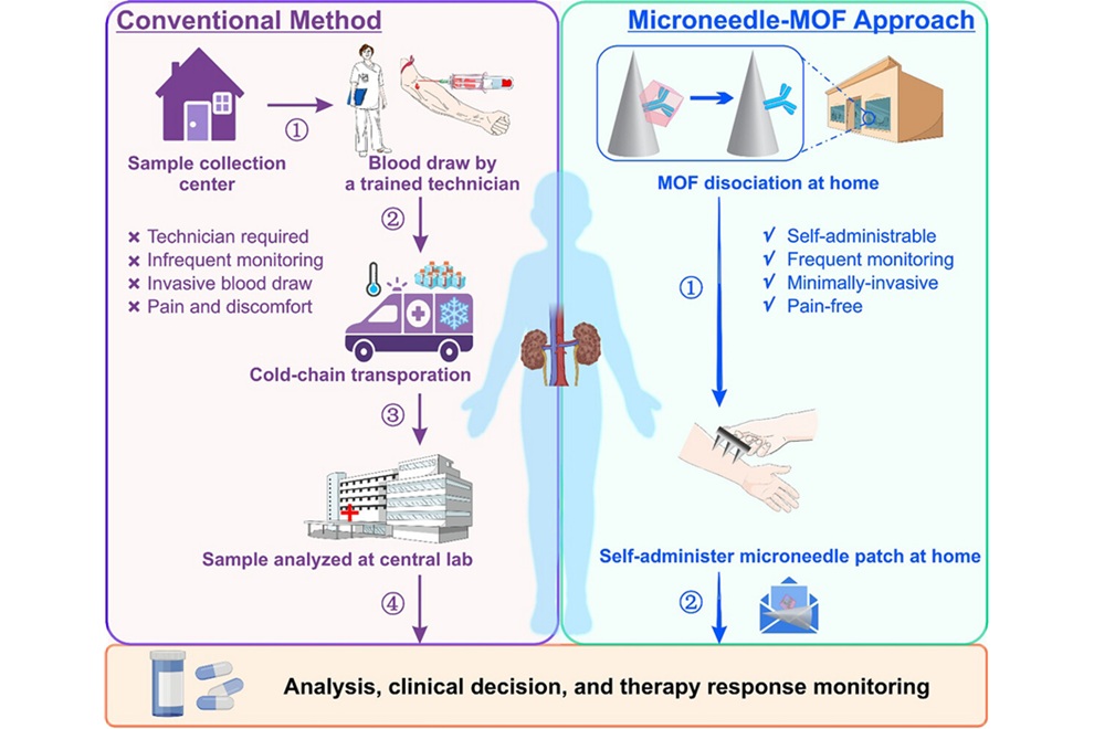
Microneedle Patch Enables At-Home Monitoring of Acute Kidney Injury
Kidney disease is often silent in its early stages, making timely detection difficult outside clinical settings. Early biomarkers such as neutrophil gelatinase–associated lipocalin can require blood draws... Read more
Business
view channel
Steris to Expand Surgical Safety Portfolio With $2.1 Billion Xodus Medical Deal
Steris has signed a definitive agreement to acquire Xodus Medical, a specialty surgical products business based in Pennsylvania, for $2.1 billion at closing. The acquisition will expand Steris’s healthcare... Read more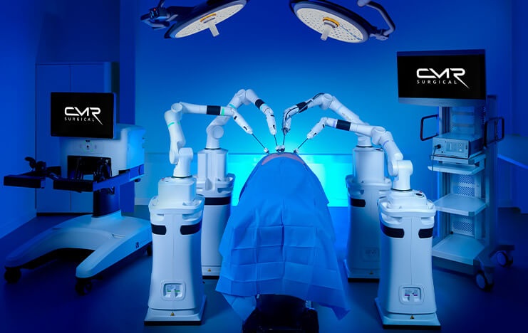
Stryker Enters Soft-Tissue Robotics Through Strategic Alliance With CMR Surgical
Stryker (Portage, MI, USA) and CMR Surgical (Cambridge, UK) have announced a strategic alliance centered on the Versius Surgical Robotic portfolio. The agreement includes an approximately USD 400 million... Read more
Terumo Neuro Acquisition Advances Liquid Embolic Technology for Chronic Subdural Hematoma
Terumo Neuro (Aliso Viejo, CA, USA) signed an agreement to acquire Arsenal Medical Inc. (Waltham, MA, USA), a privately held, clinical-stage biomaterials company. The proposed acquisition would add NeoCast,... Read more