Physicians can now excise breast lesions in their entirety, thus preserving their architectural integrity for diagnostic assessment.
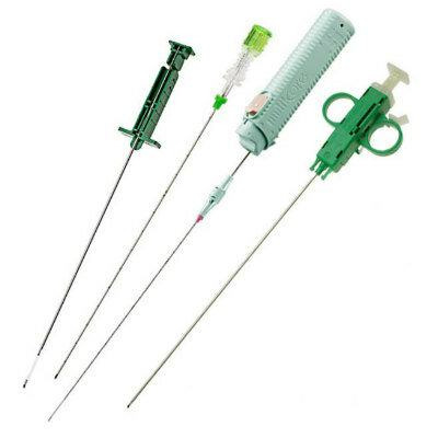
The Intact Tissue Excision System surrounds and excises an entire imaged abnormality in just ten seconds, using radiofrequency (RF) based technology. For the procedure, the surrounding tissue is numbed in five segments: four of them at 3, 6, 9, and 12 o’clock position around the lesion, and a fifth beyond it. Additional anesthesia is released along the track the wand will be used to remove the lesion. Once the area is numb, the surgeon selects a suitable diameter wand, selected according to lesion size.
Once the area is numb, the wand is inserted until it reaches the lesion position (as identified under imaging), and then envelopes it. Once captured, the wand is removed with the lesion grappled. The surgeon then leaves a marker at the excision site for future identification by X-ray to monitor the area; the lesion is then sent for biopsy. The Intact Tissue Excision System is a product of Intact Medical (Framingham, MA, USA), and has been approved by the US Food and Drug Administration (FDA) for the excision of breast lesions of up to 30 mm diameter.
“With use of Intact, physicians can offer women the option of a fast and relatively simple procedure that can remove a lesion up to 30 mm in diameter, while maintaining the lesion architecture for pathological analysis, versus capturing multiple samples of that tissue for analysis, which would not preserve architectural integrity,” said John Vacha, president and CEO of Intact Medical.
“For small breast lesions up to 30 mm in diameter, the ability of the Intact system to remove and preserve the entire lesion architecture for assessment by the pathologist combines the minimally-invasive benefits of core biopsy with the diagnostic assurances of traditional surgical excisional biopsy,” said Pat Whitworth, MD, director of the Nashville Breast Center (TN, USA). “As someone who has performed more than 1,000 procedures with the Intact, I can attest to the advantages of this option for my patients.”
Related Links:
Intact Medical
Video: Removing intact breast lesion

Gold Member
STI Test
Vivalytic Sexually Transmitted Infection (STI) Array

Gold Member
Real-Time Diagnostics Onscreen Viewer
GEMweb Live

Silver Member
Wireless Mobile ECG Recorder
NR-1207-3/NR-1207-E
New
Soft-Tissues Biopsy Needle
MR-CLEAR


")
")
")
")
")
")
")
")
")
")
")
 clearance for use with its Quantra QStat Cartridge (Photo courtesy of HemoSonics)")
 prequalification (Photo courtesy of Cepheid)")
")
")
")
")
")
")
")