3D Modeling System Accurately Predicts Pediatric Donor Heart Volumes
|
By HospiMedica International staff writers Posted on 24 Nov 2015 |
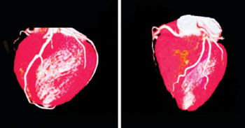
Image: 3D scan a child’s heart born with congenital heart defects (Photo courtesy of the Phoenix Children’s Hospital).
A new three dimensional (3D) computer modeling system may more accurately identify the best donor heart for a pediatric transplant patient.
To develop the new 3D system, researchers at Arizona State University (ASU; Tempe, USA) and Phoenix Children’s Hospital (AZ, USA) first created a library of 3D reconstructed hearts in healthy children weighing up to 45 kilograms, using magnetic resonance imaging (MRI) and computerized tomography (CT) scans. They then used the virtual library to predict the best donor body weight/heart size correlation needed for pediatric transplant recipients. Concomitantly, they examined before and after images from infants who had already received a heart transplant.
When the researchers compared the post-operative data from the real infants with the virtual transplant images, they found that the 3D imaging system accurately identified an appropriate size heart, validating their findings. The researchers are currently expanding the virtual library to improve prognostic capabilities, thus allowing more effective organ allocation and minimizing the number of otherwise acceptable organs that are ultimately discarded. The study was presented at the annual American Heart Association (AHA) Scientific Sessions, held during November 2015 in Orlando (FL, USA).
“It is critical to optimize the range of acceptable donors for each child. 3D reconstruction has tremendous potential to improve donor size matching,” said lead author and study presenter Jonathan Plasencia, BSc, of the ASU image processing applications lab. “We feel that we now have evidence that 3D matching can improve selection and hope this will soon help transplant doctors, patients, and their parents make the best decision by taking some of the uncertainty out of this difficult situation.”
“Analyzing future transplant cases using 3D matching will allow us to predict the true upper and lower limits of acceptable donor size. The big question is how long it will take to further test the technique and move it into actual use,” concluded Mr. Plasencia, who is a PhD student at ASU. “One day transplant teams may be able to use the 3D process to perform virtual transplants before an actual procedure to rapidly measure a donated heart to ensure a better fit and to reduce the risk of mismatching in pediatric transplants.”
Transplant centers currently assess compatibility of a potential donor heart by comparing the donor weight to the recipient weight, and then picking an upper and lower limit based on the size of the patient’s heart on chest X-ray. But the assessment is not precise and variations in size and volume can have a major effect on the recipient’s outcome.
Related Links:
Arizona State University
Phoenix Children’s Hospital
Latest Health IT News
- Machine Learning Model Improves Mortality Risk Prediction for Cardiac Surgery Patients
- Strategic Collaboration to Develop and Integrate Generative AI into Healthcare
- AI-Enabled Operating Rooms Solution Helps Hospitals Maximize Utilization and Unlock Capacity
- AI Predicts Pancreatic Cancer Three Years before Diagnosis from Patients’ Medical Records
- First Fully Autonomous Generative AI Personalized Medical Authorizations System Reduces Care Delay
- Electronic Health Records May Be Key to Improving Patient Care, Study Finds
- AI Trained for Specific Vocal Biomarkers Could Accurately Predict Coronary Artery Disease
- First-Ever AI Test for Early Diagnosis of Alzheimer’s to Be Expanded to Diagnosis of Parkinson’s Disease
- New Self-Learning AI-Based Algorithm Reads Electrocardiograms to Spot Unseen Signs of Heart Failure
- Autonomous Robot Performs COVID-19 Nasal Swab Tests

- Statistical Tool Predicts COVID-19 Peaks Worldwide
- Wireless-Controlled Soft Neural Implant Stimulates Brain Cells
- Tiny Polymer Stent Could Treat Pediatric Urethral Strictures
- Human Torso Simulator Helps Design Brace Innovations
- 3D Bioprinting Rebuilds the Human Heart
Channels
Artificial Intelligence
view channel
AI-Powered Algorithm to Revolutionize Detection of Atrial Fibrillation
Atrial fibrillation (AFib), a condition characterized by an irregular and often rapid heart rate, is linked to increased risks of stroke and heart failure. This is because the irregular heartbeat in AFib... Read more
AI Diagnostic Tool Accurately Detects Valvular Disorders Often Missed by Doctors
Doctors generally use stethoscopes to listen for the characteristic lub-dub sounds made by heart valves opening and closing. They also listen for less prominent sounds that indicate problems with these valves.... Read moreCritical Care
view channel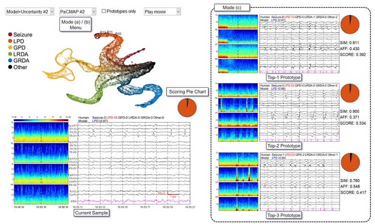
AI Doubles Medical Professionals’ Accuracy in Reading EEG Charts of ICU Patients
Electroencephalography (EEG) readings are crucial for detecting when unconscious patients may be experiencing or are at risk of seizures. EEGs involve placing small sensors on the scalp to measure the... Read moreFlexible Device Enables Sweat Gland Stimulation and Simultaneous Biosensing
Human sweat is rich in biomarkers that can be used to monitor a range of health conditions, from diabetes to genetic disorders. Many users prefer sweat sampling over blood collection because it is painless.... Read more
WHO Publishes First Global Guidelines to Reduce Bloodstream Infections from Catheter Use
Up to 70% of all inpatients require a catheter, specifically a peripherally inserted catheter (PIVC), at some point during their hospital stay. Patients who receive treatments via catheters are particularly... Read moreSurgical Techniques
view channel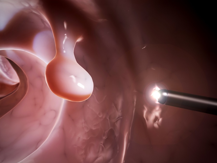
Study Warns Against Dangerous Smoke Levels Produced During Endoscopic Gastrointestinal Procedures
Healthcare professionals involved in certain smoke-generating endoscopic gastrointestinal procedures, such as those using electrical current to excise polyps, may be exposed to toxin levels comparable... Read more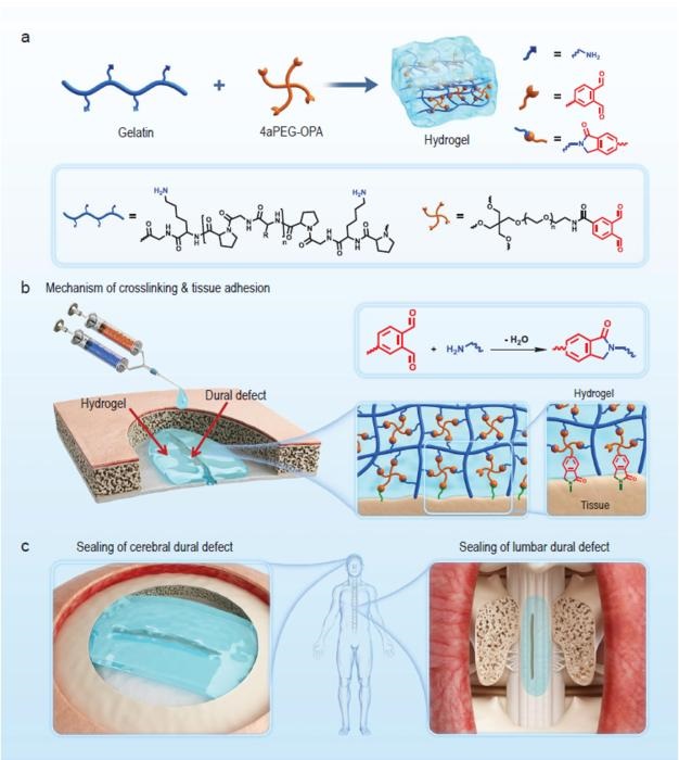
New Hydrogel Sealant Effective at Sealing Dural Defects and Preventing Postoperative Adhesion
The dura mater is a fibrous membrane of connective tissue that envelops the brain and spinal cord. In neurosurgical procedures that require access to the brain or spinal cord, opening the dura mater often... Read more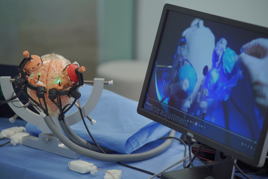
MRI-Guided Multi-Stage Robotic Positioner Enhances Stereotactic Neurosurgery Precision
Magnetic resonance imaging (MRI) offers significant benefits in neurosurgery, providing detailed 3D visualizations of neurovascular structures and tumors. Traditionally, its use has been restricted to... Read morePatient Care
view channelFirst-Of-Its-Kind Portable Germicidal Light Technology Disinfects High-Touch Clinical Surfaces in Seconds
Reducing healthcare-acquired infections (HAIs) remains a pressing issue within global healthcare systems. In the United States alone, 1.7 million patients contract HAIs annually, leading to approximately... Read more
Surgical Capacity Optimization Solution Helps Hospitals Boost OR Utilization
An innovative solution has the capability to transform surgical capacity utilization by targeting the root cause of surgical block time inefficiencies. Fujitsu Limited’s (Tokyo, Japan) Surgical Capacity... Read more
Game-Changing Innovation in Surgical Instrument Sterilization Significantly Improves OR Throughput
A groundbreaking innovation enables hospitals to significantly improve instrument processing time and throughput in operating rooms (ORs) and sterile processing departments. Turbett Surgical, Inc.... Read moreHealth IT
view channel
Machine Learning Model Improves Mortality Risk Prediction for Cardiac Surgery Patients
Machine learning algorithms have been deployed to create predictive models in various medical fields, with some demonstrating improved outcomes compared to their standard-of-care counterparts.... Read more
Strategic Collaboration to Develop and Integrate Generative AI into Healthcare
Top industry experts have underscored the immediate requirement for healthcare systems and hospitals to respond to severe cost and margin pressures. Close to half of U.S. hospitals ended 2022 in the red... Read more
AI-Enabled Operating Rooms Solution Helps Hospitals Maximize Utilization and Unlock Capacity
For healthcare organizations, optimizing operating room (OR) utilization during prime time hours is a complex challenge. Surgeons and clinics face difficulties in finding available slots for booking cases,... Read more
AI Predicts Pancreatic Cancer Three Years before Diagnosis from Patients’ Medical Records
Screening for common cancers like breast, cervix, and prostate cancer relies on relatively simple and highly effective techniques, such as mammograms, Pap smears, and blood tests. These methods have revolutionized... Read morePoint of Care
view channel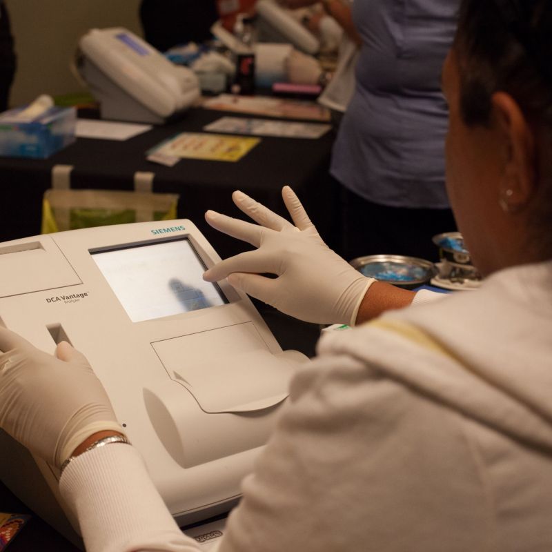
POCT for Infectious Diseases Delivers Laboratory Equivalent Pathology Results
On-site pathology tests for infectious diseases in rural and remote locations can achieve the same level of reliability and accuracy as those conducted in hospital laboratories, a recent study suggests.... Read more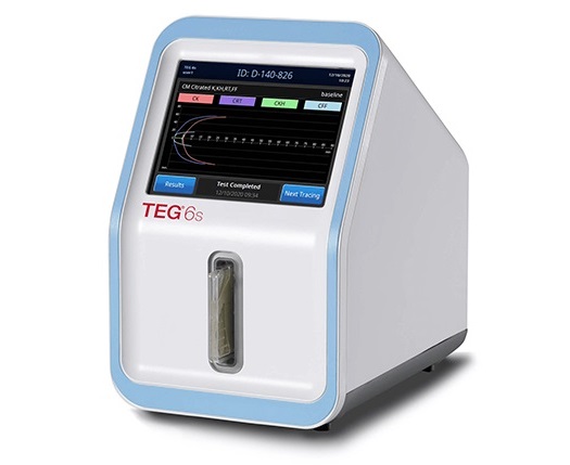
Cartridge-Based Hemostasis Analyzer System Enables Faster Coagulation Testing
Quickly assessing a patient's total hemostasis status can be critical to influencing clinical outcomes and using blood products. Haemonetics Corporation (Boston, MA, USA) has now obtained 510(k) clearance... Read more
Critical Bleeding Management System to Help Hospitals Further Standardize Viscoelastic Testing
Surgical procedures are often accompanied by significant blood loss and the subsequent high likelihood of the need for allogeneic blood transfusions. These transfusions, while critical, are linked to various... Read moreBusiness
view channel
MEDICA INNOVATION FORUM for the Healthcare Innovations of the Future
By always offering innovations and updating existing program formats, the internationally leading medical trade fair MEDICA in Düsseldorf has been successful for over half a century and always gives its... Read more


