


3D Body Mapping Helps Repair Cellular Damage
|
By HospiMedica International staff writers Posted on 02 Jul 2019 |
|
.")
Image: A floating 3D scaffold providing efficient tissue engineering monitoring (Photo courtesy of ACS Nano).
A new study reveals an innovative three-dimensional (3D) instrumented mapping technology that can monitor and track the behavior of engineered cells and tissues.
Developed by researchers at Purdue University (Lafayette, IN, USA) and Hanyang University (Seoul, Republic of Korea), the ultrabuoyant 3D scaffold remains afloat on the surface of a culture medium, providing a favorable environment for the electronic components, which remain in the air while the cells reside and grow underneath. This enables high-fidelity recording of electrical cell–substrate impedance and electrophysiological signals over long periods of time, even weeks. Currently, long-term reliable 3D monitoring is limited by the wet cell culture conditions, which are unfavorable to electronic instrument settings.
The new scaffold, on the other hand, can provide real-time monitoring of the cellular behaviors and functions, thus providing a profound impact on underlying biophysics and disease modeling. A battery of comprehensive in-vitro studies undertaken by the researchers revealed the utility of the platform as an effective tool for drug screening and tissue development following cancer treatments. They are now testing the potential of the device in stem cell therapies and the regenerative treatment of diseases. The study was published in the June 19, 2019, issue of ACS Nano.
“Tissue engineering already provides new hope for hard-to-treat disorders, and our technology brings even more possibilities. My hope is to help millions of people in need,” said senior author biomedical and mechanical engineer Chi Hwan Lee, PhD, of the Purdue College of Engineering. “This device offers an expanded set of potential options to monitor cell and tissue function after surgical transplants in diseased or damaged bodies.”
Tissue engineering, often called regenerative medicine, combines cell cultures, engineering and materials methods, and biochemical and physicochemical factors to improve or replace biological tissues. It involves the use of a tissue scaffold for the formation of new viable tissue for a medical purpose. While it was once categorized as a sub-field of biomaterials, having grown in scope and importance it can be considered as a field in its own.
Related Links:
Purdue University
Hanyang University
Gold Member
STI Test
Vivalytic Sexually Transmitted Infection (STI) Array
New
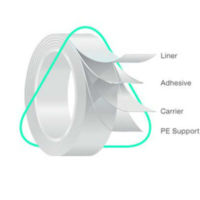
Medical Adhesive
MED 5570U
New
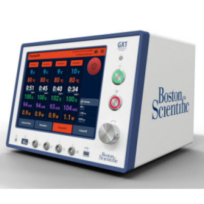
Radiofrequency Generator
GX1
Channels
Artificial Intelligence
view channelAI Analysis of Pericardial Fat Refines Long-Term Heart Disease Risk
Accurately identifying long-term cardiovascular disease risk in asymptomatic adults remains challenging for clinicians. Missed or underestimated risk delays preventive therapy and increases the chance... Read more")
Machine Learning Approach Enhances Liver Cancer Risk Stratification
Hepatocellular carcinoma, the most common form of primary liver cancer, is often detected late despite targeted surveillance programs. Current screening guidelines emphasize patients with known cirrhosis,... Read more")
, k-means++ was used to identify clusters. Three clinically distinct clusters were identified. Among those clusters, patients were divided into solid organ transplant (SOT) and non-SOT groups. Clinical characteristics and outcomes were evaluated (Masayuki Nigo et al., American Journal of Transplantation (2026). DOI: 10.1016/j.ajt.2025.10.019)")
Critical Care
view channel")
Noninvasive Monitoring Device Enables Earlier Intervention in Heart Failure
Hospitalizations for heart failure with preserved ejection fraction (HFpEF) remain common because lung congestion often worsens before symptoms prompt treatment changes. Missed early decompensation... Read more")
Automated IV Labeling Solution Improves Infusion Safety and Efficiency
Medication administration in high-acuity settings is often complicated by multiple concurrent infusions, making accurate line identification essential. In a 10-hospital intensive care unit study, 60% of... Read more)")
")
Surgical Techniques
view channel")
Ultrasound Technology Aims to Replace Invasive BPH Procedures
Benign prostatic hyperplasia (BPH) is a frequent cause of lower urinary tract symptoms in aging men and often requires invasive procedures or prolonged recovery. With prevalence expected to rise as populations... Read more")
Continuous Monitoring with Wearables Enhances Postoperative Patient Safety
Postoperative hypoxemia on general surgical wards is common and often missed by intermittent vital sign checks. Undetected low oxygen levels can delay recovery and raise the risk of complications that... Read more")
")
Patient Care
view channel")
Wearable Sleep Data Predict Adherence to Pulmonary Rehabilitation
Chronic obstructive pulmonary disease (COPD) is a long-term lung disorder that makes breathing difficult and often disturbs sleep, reducing energy for daily activities. Limited engagement in pulmonary... Read more")
Revolutionary Automatic IV-Line Flushing Device to Enhance Infusion Care
More than 80% of in-hospital patients receive intravenous (IV) therapy. Every dose of IV medicine delivered in a small volume (<250 mL) infusion bag should be followed by subsequent flushing to ensure... Read more")
")
")
Business
view channel")
")
")