3D Models Used for Research on Reptile and Bird Lungs Can Confirm Diagnosis of COVID-19 in Patients
|
By HospiMedica International staff writers Posted on 08 Sep 2020 |
Illustration
A radiologist and evolutionary anatomist from the LSU Health Sciences Center New Orleans (New Orleans, LA, USA) have found that the same techniques used for research on reptile and bird lungs can be used to help confirm the diagnosis of COVID-19 in patients.
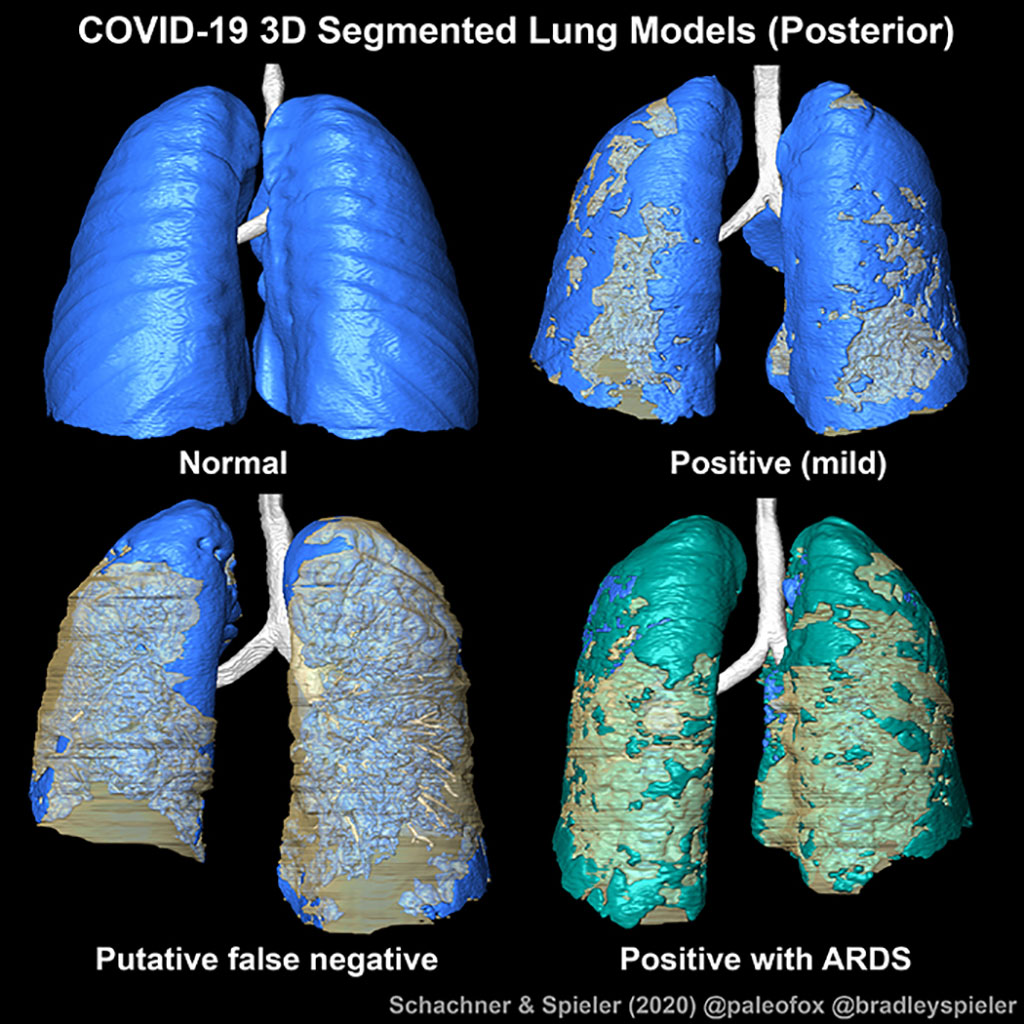
Their paper published in BMJ Case Reports demonstrates that 3D models are a strikingly clearer method for visually evaluating the distribution of COVID-19-related infection in the respiratory system. Emma R. Schachner, PhD, Associate Professor of Cell Biology & Anatomy, and Bradley Spieler, MD, Vice Chairman of Radiology Research and Associate Professor of Radiology, Internal Medicine, Urology, & Cell Biology and Anatomy at LSU Health New Orleans School of Medicine, created 3D digital models from CT scans of patients hospitalized with symptoms associated with SARS-CoV-2.
Three patients who were suspected of having COVID-19 underwent contrast enhanced thoracic CT when their symptoms worsened. Two had tested positive for SARS-CoV-2, but one was reverse transcription chain reaction (RT-PCR) negative. But because this patient had compelling clinical and imaging, the result was presumed to be a false negative. Given diagnostic challenges with respect to false negative results by RT-PCR, the gold standard for COVID-19 diagnostic screening, CT can be helpful in establishing this diagnosis. Importantly, these CT features can range in form and structure and appear to correlate with disease progression. This allows for 3D segmentation of the data in which lung tissue can be volumetrically quantified or airflow patterns could be modeled.
The CT scans were all segmented into 3D digital surface models using the scientific visualization program Avizo (Thermofisher Scientific) and techniques that the Schachner Lab uses for evolutionary anatomy research. To date, there haven’t been good models of what COVID is doing to the lungs. So, this project focused on the visualization of the lung damage in the 3D models as compared to previous methods that have been published - volume-rendered models and straight 2D screen shots of CT scans and radiographs. The three models all show varying degrees of COVID-19 related infection in the respiratory tissues - particularly along the back of the lungs, and bottom sections. They more clearly show COVID-19-related infection in the respiratory system compared to radiographs (x-rays), CT scans, or RT-PCR testing alone.
“The full effect of COVID-19 on the respiratory system remains unknown, but the 3D digital segmented models provide clinicians a new tool to evaluate the extent and distribution of the disease in one encapsulated view,” said Dr. Spieler. “This is especially useful in the case where RT-PCR for SARS-CoV-2 is negative but there is strong clinical suspicion for COVID-19.”
Related Links:
LSU Health Sciences Center New Orleans
Latest COVID-19 News
- Low-Cost System Detects SARS-CoV-2 Virus in Hospital Air Using High-Tech Bubbles
- World's First Inhalable COVID-19 Vaccine Approved in China
- COVID-19 Vaccine Patch Fights SARS-CoV-2 Variants Better than Needles
- Blood Viscosity Testing Can Predict Risk of Death in Hospitalized COVID-19 Patients
- ‘Covid Computer’ Uses AI to Detect COVID-19 from Chest CT Scans
- MRI Lung-Imaging Technique Shows Cause of Long-COVID Symptoms
- Chest CT Scans of COVID-19 Patients Could Help Distinguish Between SARS-CoV-2 Variants
- Specialized MRI Detects Lung Abnormalities in Non-Hospitalized Long COVID Patients
- AI Algorithm Identifies Hospitalized Patients at Highest Risk of Dying From COVID-19
- Sweat Sensor Detects Key Biomarkers That Provide Early Warning of COVID-19 and Flu
- Study Assesses Impact of COVID-19 on Ventilation/Perfusion Scintigraphy
- CT Imaging Study Finds Vaccination Reduces Risk of COVID-19 Associated Pulmonary Embolism
- Third Day in Hospital a ‘Tipping Point’ in Severity of COVID-19 Pneumonia
- Longer Interval Between COVID-19 Vaccines Generates Up to Nine Times as Many Antibodies
- AI Model for Monitoring COVID-19 Predicts Mortality Within First 30 Days of Admission
- AI Predicts COVID Prognosis at Near-Expert Level Based Off CT Scans
Channels
Critical Care
view channel
Mass Manufactured Nanoparticles to Deliver Cancer Drugs Directly to Tumors
Polymer-coated nanoparticles loaded with therapeutic drugs hold significant potential for treating cancers, including ovarian cancer. These particles can be precisely directed to tumors, delivering their... Read more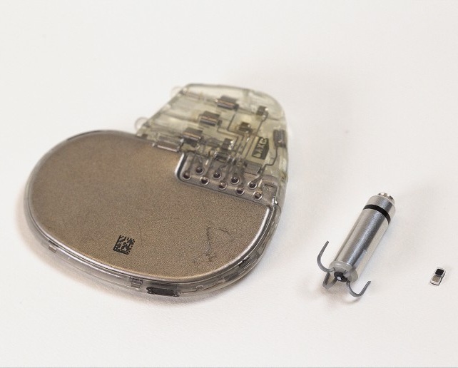
World’s Smallest Pacemaker Fits Inside Syringe Tip
After heart surgery, many patients require temporary pacemakers either to regulate the heart rate while waiting for a permanent pacemaker or to support normal heart rhythm during recovery.... Read more
AI-Powered, Internet-Connected Medical Devices to Revolutionize Healthcare, Finds Study
A new study suggests that artificial intelligence (AI)-powered, internet-connected medical devices have the potential to transform healthcare by enabling earlier detection of diseases, real-time patient... Read moreSurgical Techniques
view channel
New Transcatheter Valve Found Safe and Effective for Treating Aortic Regurgitation
Aortic regurgitation is a condition in which the aortic valve does not close properly, allowing blood to flow backward into the left ventricle. This results in decreased blood flow from the heart to the... Read more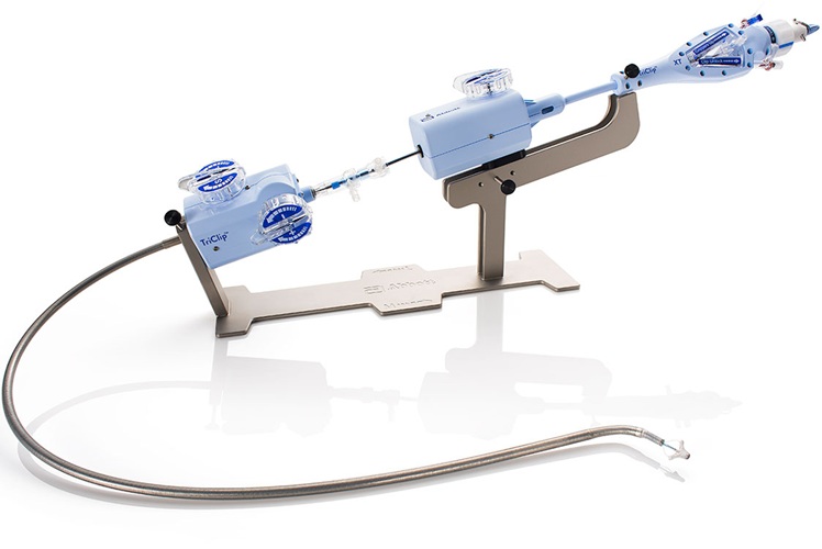
Minimally Invasive Valve Repair Reduces Hospitalizations in Severe Tricuspid Regurgitation Patients
The tricuspid valve is one of the four heart valves, responsible for regulating blood flow from the right atrium (the heart's upper-right chamber) to the right ventricle (the lower-right chamber).... Read morePatient Care
view channel
Portable Biosensor Platform to Reduce Hospital-Acquired Infections
Approximately 4 million patients in the European Union acquire healthcare-associated infections (HAIs) or nosocomial infections each year, with around 37,000 deaths directly resulting from these infections,... Read moreFirst-Of-Its-Kind Portable Germicidal Light Technology Disinfects High-Touch Clinical Surfaces in Seconds
Reducing healthcare-acquired infections (HAIs) remains a pressing issue within global healthcare systems. In the United States alone, 1.7 million patients contract HAIs annually, leading to approximately... Read more
Surgical Capacity Optimization Solution Helps Hospitals Boost OR Utilization
An innovative solution has the capability to transform surgical capacity utilization by targeting the root cause of surgical block time inefficiencies. Fujitsu Limited’s (Tokyo, Japan) Surgical Capacity... Read more
Game-Changing Innovation in Surgical Instrument Sterilization Significantly Improves OR Throughput
A groundbreaking innovation enables hospitals to significantly improve instrument processing time and throughput in operating rooms (ORs) and sterile processing departments. Turbett Surgical, Inc.... Read moreHealth IT
view channel
Printable Molecule-Selective Nanoparticles Enable Mass Production of Wearable Biosensors
The future of medicine is likely to focus on the personalization of healthcare—understanding exactly what an individual requires and delivering the appropriate combination of nutrients, metabolites, and... Read more
Smartwatches Could Detect Congestive Heart Failure
Diagnosing congestive heart failure (CHF) typically requires expensive and time-consuming imaging techniques like echocardiography, also known as cardiac ultrasound. Previously, detecting CHF by analyzing... Read morePoint of Care
view channel
Handheld, Sound-Based Diagnostic System Delivers Bedside Blood Test Results in An Hour
Patients who go to a doctor for a blood test often have to contend with a needle and syringe, followed by a long wait—sometimes hours or even days—for lab results. Scientists have been working hard to... Read moreBusiness
view channel
Expanded Collaboration to Transform OR Technology Through AI and Automation
The expansion of an existing collaboration between three leading companies aims to develop artificial intelligence (AI)-driven solutions for smart operating rooms with sophisticated monitoring and automation.... Read more


