Machine Learning Algorithm for Identifying Single Virus Particles Could Lead to More Accurate, Fast COVID-19 Test
|
By HospiMedica International staff writers Posted on 30 Nov 2020 |
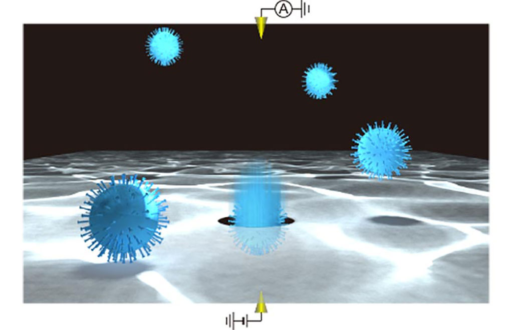
Image: Single virus particle detections using a solid-state nanopore (Photo courtesy of Osaka University)
A new system for single-virion identification of common respiratory pathogens that uses a machine learning algorithm trained on changes in current across silicon nanopores may lead to fast and accurate screening tests for diseases like COVID-19 and influenza.
Scientists at Osaka University (Suita, Japan) have introduced a new system using silicon nanopores sensitive enough to detect even a single virus particle when coupled with a machine learning algorithm. In this method, a silicon nitride layer just 50 nm thick suspended on a silicon wafer has tiny nanopores added, which are themselves only 300 nm in diameter. When a voltage difference is applied to the solution on either side of the wafer, ions travel through the nanopores in a process called electrophoresis. The motion of the ions can be monitored by the current they generate, and when a viral particle enters a nanopore, it blocks some of the ions from passing through, leading to a transient dip in current. Each dip reflects the physical properties of the particle, such as volume, surface charge, and shape, so they can be used to identify the kind of virus.
The natural variation in the physical properties of virus particles had previously hindered implementation of this approach. However, using machine learning, the team built a classification algorithm trained with signals from known viruses to determine the identity of new samples. The computer can discriminate the differences in electrical current waveforms that cannot be identified by human eyes, which enables highly accurate virus classification. In addition to coronavirus, the system was tested with similar pathogens - respiratory syncytial virus, adenovirus, influenza A, and influenza B. The team believes that coronaviruses are especially well-suited for this technique since their spiky outer proteins may even allow different strains to be classified separately. Compared with other rapid viral tests like polymerase chain reaction or antibody-based screens, the new method is much faster and does not require costly reagents, which may lead to improved diagnostic tests for emerging viral particles that cause infectious diseases such as COVID-19.
“By combining single-particle nanopore sensing with artificial intelligence, we were able to achieve highly accurate identification of multiple viral species,” said senior author Makusu Tsutsui.
“This work will help with the development of a virus test kit that outperforms conventional viral inspection methods,” added last author Tomoji Kawai.
Related Links:
Osaka University

SARS‑CoV‑2/Flu A/Flu B/RSV Sample-To-Answer Test
SARS‑CoV‑2/Flu A/Flu B/RSV Cartridge (CE-IVD)
Latest COVID-19 News
- Low-Cost System Detects SARS-CoV-2 Virus in Hospital Air Using High-Tech Bubbles
- World's First Inhalable COVID-19 Vaccine Approved in China
- COVID-19 Vaccine Patch Fights SARS-CoV-2 Variants Better than Needles
- Blood Viscosity Testing Can Predict Risk of Death in Hospitalized COVID-19 Patients
- ‘Covid Computer’ Uses AI to Detect COVID-19 from Chest CT Scans
- MRI Lung-Imaging Technique Shows Cause of Long-COVID Symptoms
- Chest CT Scans of COVID-19 Patients Could Help Distinguish Between SARS-CoV-2 Variants
- Specialized MRI Detects Lung Abnormalities in Non-Hospitalized Long COVID Patients
- AI Algorithm Identifies Hospitalized Patients at Highest Risk of Dying From COVID-19
- Sweat Sensor Detects Key Biomarkers That Provide Early Warning of COVID-19 and Flu
- Study Assesses Impact of COVID-19 on Ventilation/Perfusion Scintigraphy
- CT Imaging Study Finds Vaccination Reduces Risk of COVID-19 Associated Pulmonary Embolism
- Third Day in Hospital a ‘Tipping Point’ in Severity of COVID-19 Pneumonia
- Longer Interval Between COVID-19 Vaccines Generates Up to Nine Times as Many Antibodies
- AI Model for Monitoring COVID-19 Predicts Mortality Within First 30 Days of Admission
- AI Predicts COVID Prognosis at Near-Expert Level Based Off CT Scans
Channels
Critical Care
view channel
Novel Intrabronchial Method Delivers Cell Therapies in Critically Ill Patients on External Lung Support
Until now, administering cell therapies to patients on extracorporeal membrane oxygenation (ECMO)—a life-support system typically used for severe lung failure—has been nearly impossible.... Read more
Generative AI Technology Detects Heart Disease Earlier Than Conventional Methods
Detecting heart dysfunction early using cost-effective and widely accessible tools like electrocardiograms (ECGs) and efficiently directing the right patients for more expensive imaging tests remains a... Read more
Wearable Technology Predicts Cardiovascular Risk by Continuously Monitoring Heart Rate Recovery
The heart's response to physical activity is a vital early indicator of changes in health, particularly in cardiovascular function and mortality. Extensive research has demonstrated a connection between... Read more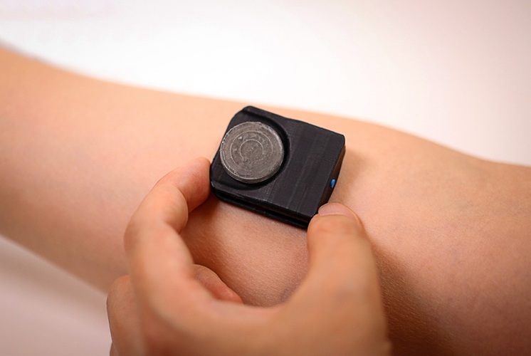
Wearable Health Monitoring Device Measures Gases Emitted from and Absorbed by Skin
The skin plays a vital role in protecting our body from external elements. A key component of this protective function is the skin barrier, which consists of tightly woven proteins and fats that help retain... Read moreSurgical Techniques
view channel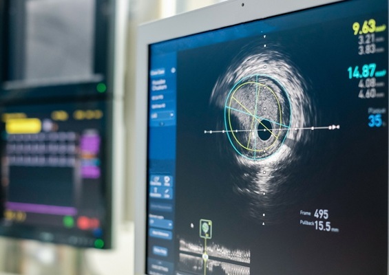
Intravascular Imaging for Guiding Stent Implantation Ensures Safer Stenting Procedures
Patients diagnosed with coronary artery disease, which is caused by plaque accumulation within the arteries leading to chest pain, shortness of breath, and potential heart attacks, frequently undergo percutaneous... Read more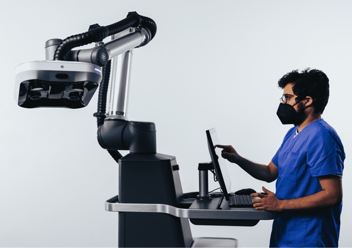
World's First AI Surgical Guidance Platform Allows Surgeons to Measure Success in Real-Time
Surgeons have always faced challenges in measuring their progress toward surgical goals during procedures. Traditionally, obtaining measurements required stepping out of the sterile environment to perform... Read morePatient Care
view channel
Portable Biosensor Platform to Reduce Hospital-Acquired Infections
Approximately 4 million patients in the European Union acquire healthcare-associated infections (HAIs) or nosocomial infections each year, with around 37,000 deaths directly resulting from these infections,... Read moreFirst-Of-Its-Kind Portable Germicidal Light Technology Disinfects High-Touch Clinical Surfaces in Seconds
Reducing healthcare-acquired infections (HAIs) remains a pressing issue within global healthcare systems. In the United States alone, 1.7 million patients contract HAIs annually, leading to approximately... Read more
Surgical Capacity Optimization Solution Helps Hospitals Boost OR Utilization
An innovative solution has the capability to transform surgical capacity utilization by targeting the root cause of surgical block time inefficiencies. Fujitsu Limited’s (Tokyo, Japan) Surgical Capacity... Read more
Game-Changing Innovation in Surgical Instrument Sterilization Significantly Improves OR Throughput
A groundbreaking innovation enables hospitals to significantly improve instrument processing time and throughput in operating rooms (ORs) and sterile processing departments. Turbett Surgical, Inc.... Read moreHealth IT
view channel
Printable Molecule-Selective Nanoparticles Enable Mass Production of Wearable Biosensors
The future of medicine is likely to focus on the personalization of healthcare—understanding exactly what an individual requires and delivering the appropriate combination of nutrients, metabolites, and... Read more
Smartwatches Could Detect Congestive Heart Failure
Diagnosing congestive heart failure (CHF) typically requires expensive and time-consuming imaging techniques like echocardiography, also known as cardiac ultrasound. Previously, detecting CHF by analyzing... Read moreBusiness
view channel
Expanded Collaboration to Transform OR Technology Through AI and Automation
The expansion of an existing collaboration between three leading companies aims to develop artificial intelligence (AI)-driven solutions for smart operating rooms with sophisticated monitoring and automation.... Read more


