Soft X-Ray Tomography 3D Scans Show How Cells Respond to SARS-CoV-2 Infection and to Possible Treatments
|
By HospiMedica International staff writers Posted on 28 Feb 2022 |
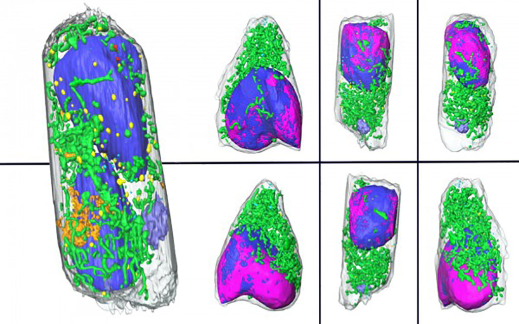
An extremely fast new 3D imaging method can show how cells respond to SARS-CoV-2 infection and to possible treatments.
Researchers from Berkeley Lab (Berkeley, CA, USA) and Heidelberg University (Heidelberg, Germany) have cranked up the speed of imaging infected cells using soft X-ray tomography (SXT), a microscopic imaging technique that can generate incredibly detailed, three-dimensional scans. Their approach takes mere minutes to gather data that would require weeks of prep and analysis with other methods, giving scientists an easy way to quickly examine how our cells’ internal machinery responds to SARS-CoV-2, or other pathogens, as well as how the cells respond to drugs designed to treat the infection.
SXT was developed at Berkeley Lab in the early 2000s to fill in the gaps left by other cellular imaging techniques and is currently offered to investigators worldwide even as the researchers continue to refine the approach. As part of a study, the researchers performed SXT on human lung cell samples. The team carefully infected the cells with SARS-CoV-2 and then chemically fixed them with aldehyde-based compounds – a process that kills cells and preserves them, immobilized, in their last living state (and also inactivates any remaining viral particles) – at six and 24 hours post-infection.
The entire team was jubilant when the resulting 3D images had the same level of exquisite detail and clarity that SXT is known for, despite the chemical fixation done to the cells. The takeaway is that their approach will allow many labs to safely image infected cells without the inherent risks – and corresponding required safety protocols – of working with live infected cells. Upon conducting the tomography sessions and image analysis, the researchers were pleasantly surprised to see how SXT captured changes to different organelles within the lung cells at very high resolution after very little time spent on sample preparation and without use of stains or labeling. These additional steps are often needed to generate cell maps wherein the different internal components are easily distinguishable.
Now that they’ve demonstrated the potential of using whole-cell SXT to safely image virus-infected cells, the researchers believe that their findings will help the global scientific community study COVID-19 and potentially other diseases. The team is already putting the technique to good use and has begun using whole-cell SXT to examine how human cells respond to several experimental COVID-19-treating drugs. They hope the rapid turnaround for results will help expedite the drug development process, getting additional effective treatments on the market sooner. They also plan to use the technology to understand the progress of infections caused by other viral agents.
“Prior to our imaging technique, if one wanted to know what was going on inside a cell, and to learn what changes had occurred upon an infection, they'd have to go through the process of fixing, slicing, and staining the cells in order to analyze them by electron microscopy. With all the steps involved, it would take weeks to get the answer. We can do it in a day,” said project co-lead Carolyn Larabell, a Berkeley Lab faculty scientist in the Biosciences Area. “So, it really speeds up the process of examining cells, the consequences to infection, and the consequences of treating a patient with a drug that may or may not cure or prevent the disease.”
Related Links:
Berkeley Lab
Heidelberg University
Latest COVID-19 News
- Low-Cost System Detects SARS-CoV-2 Virus in Hospital Air Using High-Tech Bubbles
- World's First Inhalable COVID-19 Vaccine Approved in China
- COVID-19 Vaccine Patch Fights SARS-CoV-2 Variants Better than Needles
- Blood Viscosity Testing Can Predict Risk of Death in Hospitalized COVID-19 Patients
- ‘Covid Computer’ Uses AI to Detect COVID-19 from Chest CT Scans
- MRI Lung-Imaging Technique Shows Cause of Long-COVID Symptoms
- Chest CT Scans of COVID-19 Patients Could Help Distinguish Between SARS-CoV-2 Variants
- Specialized MRI Detects Lung Abnormalities in Non-Hospitalized Long COVID Patients
- AI Algorithm Identifies Hospitalized Patients at Highest Risk of Dying From COVID-19
- Sweat Sensor Detects Key Biomarkers That Provide Early Warning of COVID-19 and Flu
- Study Assesses Impact of COVID-19 on Ventilation/Perfusion Scintigraphy
- CT Imaging Study Finds Vaccination Reduces Risk of COVID-19 Associated Pulmonary Embolism
- Third Day in Hospital a ‘Tipping Point’ in Severity of COVID-19 Pneumonia
- Longer Interval Between COVID-19 Vaccines Generates Up to Nine Times as Many Antibodies
- AI Model for Monitoring COVID-19 Predicts Mortality Within First 30 Days of Admission
- AI Predicts COVID Prognosis at Near-Expert Level Based Off CT Scans
Channels
Critical Care
view channel
Discovery of Heart’s Hidden Geometry to Revolutionize ECG Interpretation
Electrocardiograms (ECGs) are one of the most widely used tools in diagnosing heart conditions, but their accuracy can be affected by natural variations in individual heart anatomy. Traditional ECG interpretation... Read more
New Approach Improves Diagnostic Accuracy for Esophageal Motility Disorders
Esophageal motility disorders are conditions in which the muscles of the esophagus fail to function properly, often resulting in difficulty swallowing. Diagnosing these disorders can be challenging, as... Read more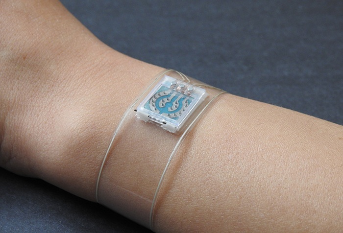
Wristband Sensor Provides All-In-One Monitoring for Diabetes and Cardiovascular Care
Diabetes management is a complex and evolving challenge, particularly when it comes to accurately tracking and understanding how various factors affect blood sugar levels and overall health.... Read more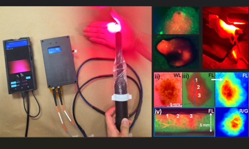
Handheld Device Enables Imaging and Treatment of Oral Cancer in Low-Resource Settings
Oral cancer is a growing public health concern, particularly in South Asia, where it affects tens of thousands of people each year. In India, oral cancer accounts for 40% of all cancers, largely driven... Read moreSurgical Techniques
view channel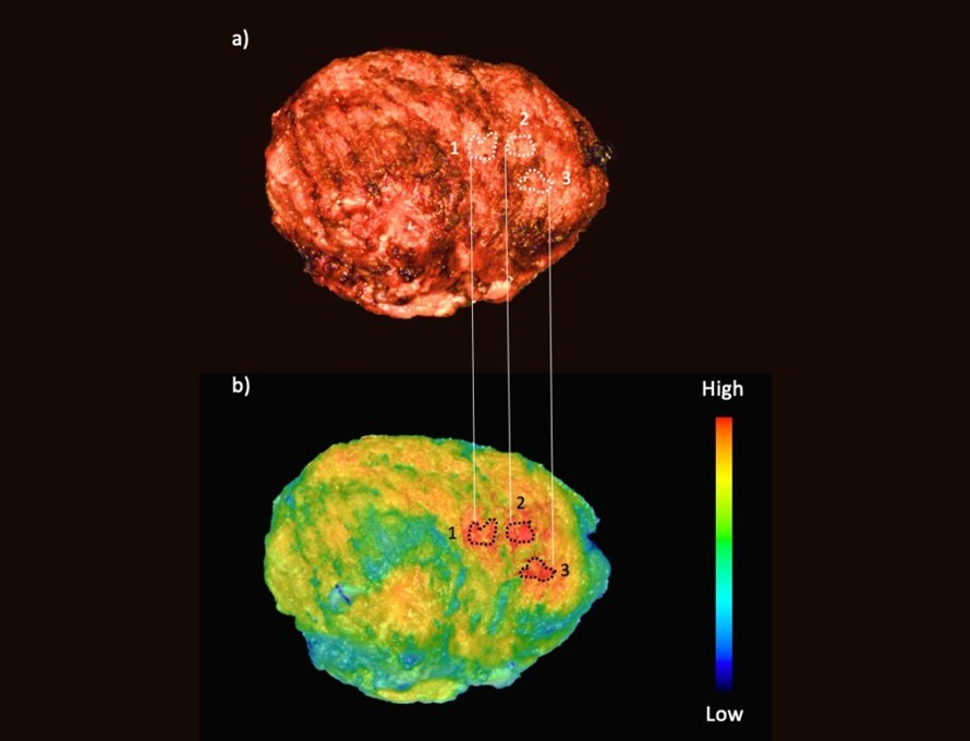
Fluorescent Imaging Agent ‘Lights Up’ Nerves for Better Visualization During Surgery
Surgical nerve injury is a significant concern in head and neck surgeries, where nerves are at risk of being inadvertently damaged during procedures. Such injuries can lead to complications that may impact... Read more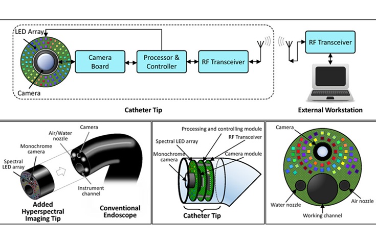
LED-Based Imaging System Could Transform Cancer Detection in Endoscopy
Gastrointestinal cancers remain one of the most common and challenging forms of cancer to diagnose accurately. Despite the widespread use of endoscopy for screening and diagnosis, the procedure still misses... Read morePatient Care
view channel
Revolutionary Automatic IV-Line Flushing Device to Enhance Infusion Care
More than 80% of in-hospital patients receive intravenous (IV) therapy. Every dose of IV medicine delivered in a small volume (<250 mL) infusion bag should be followed by subsequent flushing to ensure... Read more
VR Training Tool Combats Contamination of Portable Medical Equipment
Healthcare-associated infections (HAIs) impact one in every 31 patients, cause nearly 100,000 deaths each year, and cost USD 28.4 billion in direct medical expenses. Notably, up to 75% of these infections... Read more
Portable Biosensor Platform to Reduce Hospital-Acquired Infections
Approximately 4 million patients in the European Union acquire healthcare-associated infections (HAIs) or nosocomial infections each year, with around 37,000 deaths directly resulting from these infections,... Read moreFirst-Of-Its-Kind Portable Germicidal Light Technology Disinfects High-Touch Clinical Surfaces in Seconds
Reducing healthcare-acquired infections (HAIs) remains a pressing issue within global healthcare systems. In the United States alone, 1.7 million patients contract HAIs annually, leading to approximately... Read moreHealth IT
view channel
Printable Molecule-Selective Nanoparticles Enable Mass Production of Wearable Biosensors
The future of medicine is likely to focus on the personalization of healthcare—understanding exactly what an individual requires and delivering the appropriate combination of nutrients, metabolites, and... Read more
Smartwatches Could Detect Congestive Heart Failure
Diagnosing congestive heart failure (CHF) typically requires expensive and time-consuming imaging techniques like echocardiography, also known as cardiac ultrasound. Previously, detecting CHF by analyzing... Read moreBusiness
view channel
Bayer and Broad Institute Extend Research Collaboration to Develop New Cardiovascular Therapies
A research collaboration will focus on the joint discovery of novel therapeutic approaches based on findings in human genomics research related to cardiovascular diseases. Bayer (Berlin, Germany) and... Read more