CT Scan Study Shows Evidence of Persistent Lung Damage Long After COVID-19 Pneumonia
|
By HospiMedica International staff writers Posted on 01 Apr 2022 |
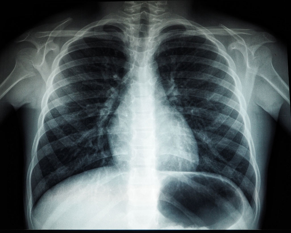
The COVID-19 pandemic, caused by the novel severe acute respiratory syndrome coronavirus type 2 (SARS-CoV-2), has considerably increased the demand for acute and post-acute healthcare worldwide. COVID-19’s short-term effects on the lungs, such as pneumonia, are well documented. Much less is known about the illness’ long-term effects on the lungs. CT has been an important imaging tool in the workup of patients suspected of having COVID-19. Now, a new study has found that some people recovering from COVID-19 pneumonia have CT evidence of damage to their lungs that persists a full year after the onset of symptoms. The study underscores radiology’s role in helping identify patients at risk for post-COVID-19 consequences and assisting in COVID-19 follow-up management.
As part of an observational study on the development of lung disease in patients with SARS-CoV-2 infection, researchers at Innsbruck Medical University (Innsbruck, Austria) looked at patterns and rates of improvement of chest CT abnormalities in patients one year after COVID-19 pneumonia. The researchers assessed lung abnormalities on chest CT in 91 participants, mean age 59 years, at several points over one year after the onset of COVID-19 symptoms.
At one year, CT abnormalities were present in 49, or 54%, of the 91 participants. Of these 49 participants, two (4%) had received outpatient treatment only, while 25 (51%) were treated on a general hospital ward and 22 (45%) had received intensive care unit (ICU) treatment. While CT abnormalities decreased in initial follow-ups, 63% of participants with abnormalities did not show any further improvement after six months. Age over 60 years, critical COVID-19 severity and male gender were associated with persistent CT abnormalities at one year. Evidence from the SARS-CoV-1 outbreak of 2002 to 2004 shows that lung abnormalities may remain detectable even after decades, but do not show any progression, according to the researchers. Recent studies, though, have shown a risk of progression of lung abnormalities such as the ones depicted on CT. The researchers intend to continue gathering data on patients with persistent CT abnormalities
“The observed chest CT abnormalities from our study are indicative of damaged lung tissue,” said study co-author Anna Luger, M.D., from the Department of Radiology at Innsbruck Medical University in Innsbruck, Austria. “However, it is currently unclear if they represent persistent scarring, and whether they regress over time or lead to pulmonary fibrosis.”
“In a recently published clinical study of our CovILD interdisciplinary working group, we were able to show that the severity of acute COVID-19, protracted systemic inflammation and the presence of residual chest CT abnormalities are strongly related to persistently impaired lung function and clinical symptoms,” said study co-author Christoph Schwabl, M.D., from Innsbruck Medical University.
“In the end, long-term follow-up, both clinical and radiological, is necessary to gather more information about the course and clinical role of persisting SARS-CoV-2 related chest CT abnormalities,” said study senior author Gerlig Widmann, M.D., chief thoracic radiologist at Innsbruck Medical University.
Related Links:
Innsbruck Medical University
Latest COVID-19 News
- Low-Cost System Detects SARS-CoV-2 Virus in Hospital Air Using High-Tech Bubbles
- World's First Inhalable COVID-19 Vaccine Approved in China
- COVID-19 Vaccine Patch Fights SARS-CoV-2 Variants Better than Needles
- Blood Viscosity Testing Can Predict Risk of Death in Hospitalized COVID-19 Patients
- ‘Covid Computer’ Uses AI to Detect COVID-19 from Chest CT Scans
- MRI Lung-Imaging Technique Shows Cause of Long-COVID Symptoms
- Chest CT Scans of COVID-19 Patients Could Help Distinguish Between SARS-CoV-2 Variants
- Specialized MRI Detects Lung Abnormalities in Non-Hospitalized Long COVID Patients
- AI Algorithm Identifies Hospitalized Patients at Highest Risk of Dying From COVID-19
- Sweat Sensor Detects Key Biomarkers That Provide Early Warning of COVID-19 and Flu
- Study Assesses Impact of COVID-19 on Ventilation/Perfusion Scintigraphy
- CT Imaging Study Finds Vaccination Reduces Risk of COVID-19 Associated Pulmonary Embolism
- Third Day in Hospital a ‘Tipping Point’ in Severity of COVID-19 Pneumonia
- Longer Interval Between COVID-19 Vaccines Generates Up to Nine Times as Many Antibodies
- AI Model for Monitoring COVID-19 Predicts Mortality Within First 30 Days of Admission
- AI Predicts COVID Prognosis at Near-Expert Level Based Off CT Scans
Channels
Critical Care
view channel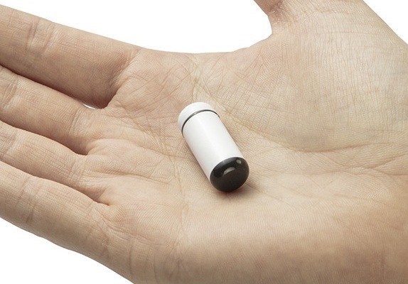
Ingestible Smart Capsule for Chemical Sensing in the Gut Moves Closer to Market
Intestinal gases are associated with several health conditions, including colon cancer, irritable bowel syndrome, and inflammatory bowel disease, and they have the potential to serve as crucial biomarkers... Read moreNovel Cannula Delivery System Enables Targeted Delivery of Imaging Agents and Drugs
Multiphoton microscopy has become an invaluable tool in neuroscience, allowing researchers to observe brain activity in real time with high-resolution imaging. A crucial aspect of many multiphoton microscopy... Read more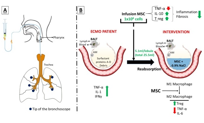
Novel Intrabronchial Method Delivers Cell Therapies in Critically Ill Patients on External Lung Support
Until now, administering cell therapies to patients on extracorporeal membrane oxygenation (ECMO)—a life-support system typically used for severe lung failure—has been nearly impossible.... Read moreSurgical Techniques
view channel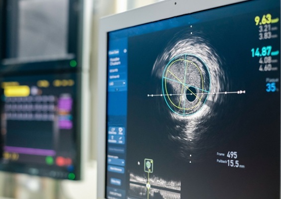
Intravascular Imaging for Guiding Stent Implantation Ensures Safer Stenting Procedures
Patients diagnosed with coronary artery disease, which is caused by plaque accumulation within the arteries leading to chest pain, shortness of breath, and potential heart attacks, frequently undergo percutaneous... Read more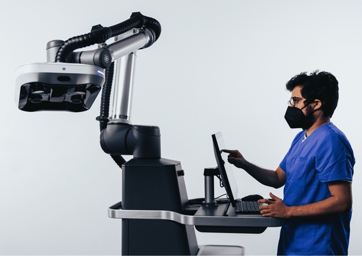
World's First AI Surgical Guidance Platform Allows Surgeons to Measure Success in Real-Time
Surgeons have always faced challenges in measuring their progress toward surgical goals during procedures. Traditionally, obtaining measurements required stepping out of the sterile environment to perform... Read morePatient Care
view channel
Portable Biosensor Platform to Reduce Hospital-Acquired Infections
Approximately 4 million patients in the European Union acquire healthcare-associated infections (HAIs) or nosocomial infections each year, with around 37,000 deaths directly resulting from these infections,... Read moreFirst-Of-Its-Kind Portable Germicidal Light Technology Disinfects High-Touch Clinical Surfaces in Seconds
Reducing healthcare-acquired infections (HAIs) remains a pressing issue within global healthcare systems. In the United States alone, 1.7 million patients contract HAIs annually, leading to approximately... Read more
Surgical Capacity Optimization Solution Helps Hospitals Boost OR Utilization
An innovative solution has the capability to transform surgical capacity utilization by targeting the root cause of surgical block time inefficiencies. Fujitsu Limited’s (Tokyo, Japan) Surgical Capacity... Read more
Game-Changing Innovation in Surgical Instrument Sterilization Significantly Improves OR Throughput
A groundbreaking innovation enables hospitals to significantly improve instrument processing time and throughput in operating rooms (ORs) and sterile processing departments. Turbett Surgical, Inc.... Read moreHealth IT
view channel
Printable Molecule-Selective Nanoparticles Enable Mass Production of Wearable Biosensors
The future of medicine is likely to focus on the personalization of healthcare—understanding exactly what an individual requires and delivering the appropriate combination of nutrients, metabolites, and... Read more
Smartwatches Could Detect Congestive Heart Failure
Diagnosing congestive heart failure (CHF) typically requires expensive and time-consuming imaging techniques like echocardiography, also known as cardiac ultrasound. Previously, detecting CHF by analyzing... Read moreBusiness
view channel
Expanded Collaboration to Transform OR Technology Through AI and Automation
The expansion of an existing collaboration between three leading companies aims to develop artificial intelligence (AI)-driven solutions for smart operating rooms with sophisticated monitoring and automation.... Read more


