


Pathology Device Averts Repeated Breast Cancer Surgeries
|
By HospiMedica International staff writers Posted on 16 Jul 2013 |
|
.")
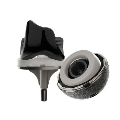
Image: The prototype device applies an adhesive film to the breast tissue before examination (Photo courtesy of Will Kirk/homewoodphoto.jhu.edu).
A new tool will allow pathologists to quickly inspect excised breast tissue while the patient is still in the operating room (OR), reducing the need for a second surgery.
Developed by graduate students at Johns Hopkins University (Baltimore, MD, USA), the prototype device applies an adhesive film to the specimen breast tissue before it is sliced; the film thus holds the delicate tissue together, preventing damage to the samples during the preparation process. The result is a sample that can be clearly reviewed by a pathologist within 20 minutes of its removal, potentially eliminating the need for a second operation on another day. The low-cost system includes a reusable applicator and a proprietary disposable film.
“We spoke to breast cancer surgeons,” said Hector Neira, MSc, one of the student inventors. “They told us that they are desperate for something that will allow them to remove the tumor in its entirety the first time, so that the patient doesn’t have to come back for a second surgery.”
“I think the students have been incredibly creative in their development of this concept, and they are addressing a very real need in the field of breast cancer surgery,” added professor of Surgery Melissa Camp, who worked with the students. “Accurate assessment of margin status during the initial operation will lead to fewer reoperations, and this will be beneficial for patients in many respects. I look forward to their continued work!”
When most tumors are removed, pathologist can quickly flash-freeze the tissue and slice off paper-thin samples for microscopic examination; if the pathologist sees that cancer cells extend to the outer edge or margin of a sample, the surgeon is advised to remove more tissue from the patient. Breast tissue, however, poses a problem; it has a high fat content and does not freeze well, causing the samples to smear, form gaps, and become unsuitable for a quick review.
Related Links:
Johns Hopkins University
Gold Member
SARS‑CoV‑2/Flu A/Flu B/RSV Sample-To-Answer Test
SARS‑CoV‑2/Flu A/Flu B/RSV Cartridge (CE-IVD)
Gold Member
Solid State Kv/Dose Multi-Sensor
AGMS-DM+
Silver Member
Wireless Mobile ECG Recorder
NR-1207-3/NR-1207-E
New
Oxidized Zirconium Implant Material
OXINIUM
Latest Surgical Techniques News
- Miniaturized Implantable Multi-Sensors Device to Monitor Vessels Health
- Tiny Robots Made Out Of Carbon Could Conduct Colonoscopy, Pelvic Exam or Blood Test
- Miniaturized Ultrasonic Scalpel Enables Faster and Safer Robotic-Assisted Surgery
- AI Assisted Reading Tool for Small Bowel Video Capsule Endoscopy Detects More Lesions
- First-Ever Contact Force Pulsed Field Ablation System to Transform Treatment of Ventricular Arrhythmias
- Caterpillar Robot with Built-In Steering System Crawls Easily Through Loops and Bends
- Tiny Wraparound Electronic Implants to Revolutionize Treatment of Spinal Cord Injuries
- Small, Implantable Cardiac Pump to Help Children Awaiting Heart Transplant
- Gastrointestinal Imaging Capsule a Game-Changer in Esophagus Surveillance and Treatment
- World’s Smallest Laser Probe for Brain Procedures Facilitates Ablation of Full Range of Targets
- Artificial Intelligence Broadens Diagnostic Abilities of Conventional Coronary Angiography
- AI-Powered Surgical Visualization Tool Supports Surgeons' Visual Recognition in Real Time
- Cutting-Edge Robotic Bronchial Endoscopic System Provides Prompt Intervention during Emergencies
- Handheld Device for Fluorescence-Guided Surgery a Game Changer for Removal of High-Grade Glioma Brain Tumors
- Porous Gel Sponge Facilitates Rapid Hemostasis and Wound Healing
- Novel Rigid Endoscope System Enables Deep Tissue Imaging During Surgery
Channels
Artificial Intelligence
view channel")
AI-Powered Algorithm to Revolutionize Detection of Atrial Fibrillation
Atrial fibrillation (AFib), a condition characterized by an irregular and often rapid heart rate, is linked to increased risks of stroke and heart failure. This is because the irregular heartbeat in AFib... Read more")
AI Diagnostic Tool Accurately Detects Valvular Disorders Often Missed by Doctors
Doctors generally use stethoscopes to listen for the characteristic lub-dub sounds made by heart valves opening and closing. They also listen for less prominent sounds that indicate problems with these valves.... Read more")
Critical Care
view channel")
Powerful AI Risk Assessment Tool Predicts Outcomes in Heart Failure Patients
Heart failure is a serious condition where the heart cannot pump sufficient blood to meet the body's needs, leading to symptoms like fatigue, weakness, and swelling in the legs and feet, and it can ultimately... Read more")
Peptide-Based Hydrogels Repair Damaged Organs and Tissues On-The-Spot
Scientists have ingeniously combined biomedical expertise with nature-inspired engineering to develop a jelly-like material that holds significant promise for immediate repairs to a wide variety of damaged... Read more")
One-Hour Endoscopic Procedure Could Eliminate Need for Insulin for Type 2 Diabetes
Over 37 million Americans are diagnosed with diabetes, and more than 90% of these cases are Type 2 diabetes. This form of diabetes is most commonly seen in individuals over 45, though an increasing number... Read morePatient Care
view channelFirst-Of-Its-Kind Portable Germicidal Light Technology Disinfects High-Touch Clinical Surfaces in Seconds
Reducing healthcare-acquired infections (HAIs) remains a pressing issue within global healthcare systems. In the United States alone, 1.7 million patients contract HAIs annually, leading to approximately... Read more")
Surgical Capacity Optimization Solution Helps Hospitals Boost OR Utilization
An innovative solution has the capability to transform surgical capacity utilization by targeting the root cause of surgical block time inefficiencies. Fujitsu Limited’s (Tokyo, Japan) Surgical Capacity... Read more")
Game-Changing Innovation in Surgical Instrument Sterilization Significantly Improves OR Throughput
A groundbreaking innovation enables hospitals to significantly improve instrument processing time and throughput in operating rooms (ORs) and sterile processing departments. Turbett Surgical, Inc.... Read more")
Health IT
view channel")
Machine Learning Model Improves Mortality Risk Prediction for Cardiac Surgery Patients
Machine learning algorithms have been deployed to create predictive models in various medical fields, with some demonstrating improved outcomes compared to their standard-of-care counterparts.... Read more")
Strategic Collaboration to Develop and Integrate Generative AI into Healthcare
Top industry experts have underscored the immediate requirement for healthcare systems and hospitals to respond to severe cost and margin pressures. Close to half of U.S. hospitals ended 2022 in the red... Read more")
AI-Enabled Operating Rooms Solution Helps Hospitals Maximize Utilization and Unlock Capacity
For healthcare organizations, optimizing operating room (OR) utilization during prime time hours is a complex challenge. Surgeons and clinics face difficulties in finding available slots for booking cases,... Read more")
AI Predicts Pancreatic Cancer Three Years before Diagnosis from Patients’ Medical Records
Screening for common cancers like breast, cervix, and prostate cancer relies on relatively simple and highly effective techniques, such as mammograms, Pap smears, and blood tests. These methods have revolutionized... Read morePoint of Care
view channel clearance for use with its Quantra QStat Cartridge (Photo courtesy of HemoSonics)")
Critical Bleeding Management System to Help Hospitals Further Standardize Viscoelastic Testing
Surgical procedures are often accompanied by significant blood loss and the subsequent high likelihood of the need for allogeneic blood transfusions. These transfusions, while critical, are linked to various... Read more prequalification (Photo courtesy of Cepheid)")
Point of Care HIV Test Enables Early Infection Diagnosis for Infants
Early diagnosis and initiation of treatment are crucial for the survival of infants infected with HIV (human immunodeficiency virus). Without treatment, approximately 50% of infants who acquire HIV during... Read more")
Whole Blood Rapid Test Aids Assessment of Concussion at Patient's Bedside
In the United States annually, approximately five million individuals seek emergency department care for traumatic brain injuries (TBIs), yet over half of those suspecting a concussion may never get it checked.... Read more")
New Generation Glucose Hospital Meter System Ensures Accurate, Interference-Free and Safe Use
A new generation glucose hospital meter system now comes with several features that make hospital glucose testing easier and more secure while continuing to offer accuracy, freedom from interference, and... Read moreBusiness
view channel")
Johnson & Johnson Acquires Cardiovascular Medical Device Company Shockwave Medical
Johnson & Johnson (New Brunswick, N.J., USA) and Shockwave Medical (Santa Clara, CA, USA) have entered into a definitive agreement under which Johnson & Johnson will acquire all of Shockwave’s... Read more")
")