


Interbody Fusion Device Provides Integrated Fixation
|
By HospiMedica International staff writers Posted on 24 Jun 2018 |
|
.")
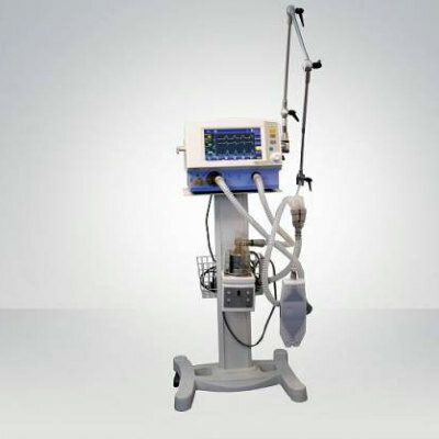
Image: Multiple views of the ENZA-A Titanium ALIF system (Photo courtesy of Camber Spine).
A minimally invasive anterior lumbar interbody fusion (ALIF) system uses autogenous bone grafts to stabilize patients with degenerative disc disease (DDD).
The Camber Spine (Wayne, PA, USA) ENZA-A Titanium ALIF system consists of a three-dimensional (3D) printed titanium body with multiple openings to allow a large volume of autogenous bone graft to be easily packed into the implant, and roughened cranial and caudal surfaces that encourage bone growth onto the surface of the device. The surfaces are deliberately designed with pores that average 500 microns in diameter, the optimal environment for bone growth that fully incorporates the implant with the vertebral bodies.
The ENZA-A features two sharpened anchor plates housed within the 3D-printed body until they are deployed into the adjacent vertebrae for fixation. Surgery time is reduced thanks to the single, inline instrumentation used to insert the device, deploy the anchor plates, and lock it in place. Patient safety is increased by minimizing the size of the incision and retraction required for implantation. The ENZA-A has been approved by the U.S. Food and Drug Administration (FDA) for use at one or two contiguous levels from L2 to S1 using supplementary fixation systems.
“With ergonomic instrumentation, this system is easy to use and makes implantation more streamlined,” said Seth Anderson, executive VP of new business development and surgeon relations at Camber Spine. “The ENZA-A is the second device in the ENZA-line of implants; this interbody, coupled with additional product launches expected later this year in the cervical and lateral markets, will continue to grow Camber Spine's presence as a market leader and innovator in minimally invasive spine surgery technology advancements.”
Interbody devices are designed to replace the intervertebral disc of the spine, enhancing stability in the region while the spine fuses. Over time, the packed bone graft material is gradually replaced by natural bone forming a solid piece. Fusion procedures typically use a posterior fixation device to the associated level, since the surgeons will implant interbody devices from an anterior approach and flip the patient over to implant a posterior pedicle screw device. This combination increases fusion success.
Related Links:
Camber Spine
Gold Member
Solid State Kv/Dose Multi-Sensor
AGMS-DM+
Gold Member
Real-Time Diagnostics Onscreen Viewer
GEMweb Live
Silver Member
Wireless Mobile ECG Recorder
NR-1207-3/NR-1207-E
New
Ventilator
TRventi-3D
Latest Surgical Techniques News
- Novel Rigid Endoscope System Enables Deep Tissue Imaging During Surgery
- Robotic Nerve ‘Cuffs’ Could Treat Various Neurological Conditions
- Flexible Microdisplay Visualizes Brain Activity in Real-Time To Guide Neurosurgeons

- Next-Gen Computer Assisted Vacuum Thrombectomy Technology Rapidly Removes Blood Clots
- Hydrogel-Based Miniaturized Electric Generators to Power Biomedical Devices
- Custom 3D-Printed Orthopedic Implants Transform Joint Replacement Surgery
- Wearable Technology Monitors and Analyzes Surgeons' Posture during Long Surgical Procedures
- Cutting-Edge Imaging Platform Detects Residual Breast Cancer Missed During Lumpectomy Surgery
- Computational Models Predict Heart Valve Leakage in Children
- Breakthrough Device Enables Clear and Real-Time Visual Guidance for Effective Cardiovascular Interventions
- World’s First Microscopic Probe to Revolutionize Early Cancer Diagnosis
- World’s Smallest Implantable Brain Stimulator Demonstrated in Human Patient
- Robotically Assisted Lung Transplants Could Soon Become a Reality
- AI to Provide Heart Transplant Surgeons with New Decision-Making Data
- New Surgical Tool Empowers Precision and Confidence in Operating Room
- Future Muscle-Powered Surgical Robots Could Perform Minimally Invasive Procedures inside Body
Channels
Artificial Intelligence
view channel")
AI-Powered Algorithm to Revolutionize Detection of Atrial Fibrillation
Atrial fibrillation (AFib), a condition characterized by an irregular and often rapid heart rate, is linked to increased risks of stroke and heart failure. This is because the irregular heartbeat in AFib... Read more")
AI Diagnostic Tool Accurately Detects Valvular Disorders Often Missed by Doctors
Doctors generally use stethoscopes to listen for the characteristic lub-dub sounds made by heart valves opening and closing. They also listen for less prominent sounds that indicate problems with these valves.... Read more")
")
Critical Care
view channel")
On-Skin Wearable Bioelectronic Device Paves Way for Intelligent Implants
A team of researchers at the University of Missouri (Columbia, MO, USA) has achieved a milestone in developing a state-of-the-art on-skin wearable bioelectronic device. This development comes from a lab... Read more")
First-Of-Its-Kind Dissolvable Stent to Improve Outcomes for Patients with Severe PAD
Peripheral artery disease (PAD) affects millions and presents serious health risks, particularly its severe form, chronic limb-threatening ischemia (CLTI). CLTI develops when arteries are blocked by plaque,... Read more")
AI Brain-Age Estimation Technology Uses EEG Scans to Screen for Degenerative Diseases
As individuals age, so do their brains. Premature aging of the brain can lead to age-related conditions such as mild cognitive impairment, dementia, or Parkinson's disease. The ability to determine "brain... Read more")
Patient Care
view channelFirst-Of-Its-Kind Portable Germicidal Light Technology Disinfects High-Touch Clinical Surfaces in Seconds
Reducing healthcare-acquired infections (HAIs) remains a pressing issue within global healthcare systems. In the United States alone, 1.7 million patients contract HAIs annually, leading to approximately... Read more")
Surgical Capacity Optimization Solution Helps Hospitals Boost OR Utilization
An innovative solution has the capability to transform surgical capacity utilization by targeting the root cause of surgical block time inefficiencies. Fujitsu Limited’s (Tokyo, Japan) Surgical Capacity... Read more")
Game-Changing Innovation in Surgical Instrument Sterilization Significantly Improves OR Throughput
A groundbreaking innovation enables hospitals to significantly improve instrument processing time and throughput in operating rooms (ORs) and sterile processing departments. Turbett Surgical, Inc.... Read more")
Health IT
view channel")
Machine Learning Model Improves Mortality Risk Prediction for Cardiac Surgery Patients
Machine learning algorithms have been deployed to create predictive models in various medical fields, with some demonstrating improved outcomes compared to their standard-of-care counterparts.... Read more")
Strategic Collaboration to Develop and Integrate Generative AI into Healthcare
Top industry experts have underscored the immediate requirement for healthcare systems and hospitals to respond to severe cost and margin pressures. Close to half of U.S. hospitals ended 2022 in the red... Read more")
AI-Enabled Operating Rooms Solution Helps Hospitals Maximize Utilization and Unlock Capacity
For healthcare organizations, optimizing operating room (OR) utilization during prime time hours is a complex challenge. Surgeons and clinics face difficulties in finding available slots for booking cases,... Read more")
AI Predicts Pancreatic Cancer Three Years before Diagnosis from Patients’ Medical Records
Screening for common cancers like breast, cervix, and prostate cancer relies on relatively simple and highly effective techniques, such as mammograms, Pap smears, and blood tests. These methods have revolutionized... Read morePoint of Care
view channel clearance for use with its Quantra QStat Cartridge (Photo courtesy of HemoSonics)")
Critical Bleeding Management System to Help Hospitals Further Standardize Viscoelastic Testing
Surgical procedures are often accompanied by significant blood loss and the subsequent high likelihood of the need for allogeneic blood transfusions. These transfusions, while critical, are linked to various... Read more prequalification (Photo courtesy of Cepheid)")
Point of Care HIV Test Enables Early Infection Diagnosis for Infants
Early diagnosis and initiation of treatment are crucial for the survival of infants infected with HIV (human immunodeficiency virus). Without treatment, approximately 50% of infants who acquire HIV during... Read more")
Whole Blood Rapid Test Aids Assessment of Concussion at Patient's Bedside
In the United States annually, approximately five million individuals seek emergency department care for traumatic brain injuries (TBIs), yet over half of those suspecting a concussion may never get it checked.... Read more")
New Generation Glucose Hospital Meter System Ensures Accurate, Interference-Free and Safe Use
A new generation glucose hospital meter system now comes with several features that make hospital glucose testing easier and more secure while continuing to offer accuracy, freedom from interference, and... Read moreBusiness
view channel")
Johnson & Johnson Acquires Cardiovascular Medical Device Company Shockwave Medical
Johnson & Johnson (New Brunswick, N.J., USA) and Shockwave Medical (Santa Clara, CA, USA) have entered into a definitive agreement under which Johnson & Johnson will acquire all of Shockwave’s... Read more")
")