Artificial Intelligence Helps Detect Rare Diseases
|
By HospiMedica International staff writers Posted on 18 Jun 2019 |

A child with Kabuki Syndrome (Photo courtesy of Wikimedia).
A new study suggests that an artificial intelligence (AI) neural network can be used to automatically combine portrait photos and genetic data to diagnose rare diseases more efficiently.
The prioritization of exome data by image analysis (PEDIA) project, under development by the University of Bonn (Germany), GeneTalk (Bonn, Germany), Charité University Medicine (Charité; Berlin, Germany), and other institutions, is designed to interpret exome data by analyzing sequence variants in portrait photographs, and integrating the results using the DeepGestalt phenotyping tool, a product of FDNA (Herzliya, Israel), which was trained with around 30,000 portrait pictures of people affected by rare syndromal diseases.
In a proof of concept study, the researchers measured the value added by computer-assisted image analysis to the diagnostic yield on a cohort consisting of 679 individuals with 105 different monogenic disorders. For each separate case, frontal photos, clinical features, and the disease-causing variants were submitted. The results showed that computer-assisted analysis of frontal photos improved the top 1% accuracy rate by more than 20–89%, and the top 10% accuracy rate by more than 5–99% for the disease-causing gene. The study was published on June 5, 2019, in Nature Genetics in Medicine.
“In combination with facial analysis, it is possible to filter out the decisive genetic factors and prioritize genes. Merging data in the neuronal network reduces data analysis time and leads to a higher rate of diagnosis,” said senior author Professor Peter Krawitz, MD, PhD, director of the Institute for Genomic Statistics and Bioinformatics at the University of Bonn. “This is of great scientific interest to us and also enables us to find a cause in some unsolved cases.”
“PEDIA is a unique example of next-generation phenotyping technologies,” said Dekel Gelbman, CEO of FDNA. “Integrating an advanced AI and facial analysis framework such as DeepGestalt into the variant analysis workflow will result in a new paradigm for superior genetic testing.”
Many rare diseases cause characteristic abnormal facial features in those affected, such as Kabuki syndrome, which is reminiscent of the make-up of a traditional Japanese form of theatre. The eyebrows are arched, the eye-distance is wide and the spaces between the eyelids are long. Another example is mucopolysaccharidosis, which leads to bone deformation, stunted growth, and learning difficulties. Such phenotype information has so far only been accessible for bioinformatics workflows after encoding into clinical terms by expert dysmorphologists.
Related Links:
University of Bonn
GeneTalk
Charité University Medicine
FDNA
Latest AI News
- AI-Powered Algorithm to Revolutionize Detection of Atrial Fibrillation
- AI Diagnostic Tool Accurately Detects Valvular Disorders Often Missed by Doctors
- New Model Predicts 10 Year Breast Cancer Risk
- AI Tool Accurately Predicts Cancer Three Years Prior to Diagnosis
- Ground-Breaking Tool Predicts 10-Year Risk of Esophageal Cancer
- AI Tool Analyzes Capsule Endoscopy Videos for Accurately Predicting Patient Outcomes for Crohn’s Disease
Channels
Critical Care
view channel
Stretchable Microneedles to Help In Accurate Tracking of Abnormalities and Identifying Rapid Treatment
The field of personalized medicine is transforming rapidly, with advancements like wearable devices and home testing kits making it increasingly easy to monitor a wide range of health metrics, from heart... Read more
Machine Learning Tool Identifies Rare, Undiagnosed Immune Disorders from Patient EHRs
Patients suffering from rare diseases often endure extensive delays in receiving accurate diagnoses and treatments, which can lead to unnecessary tests, worsening health, psychological strain, and significant... Read more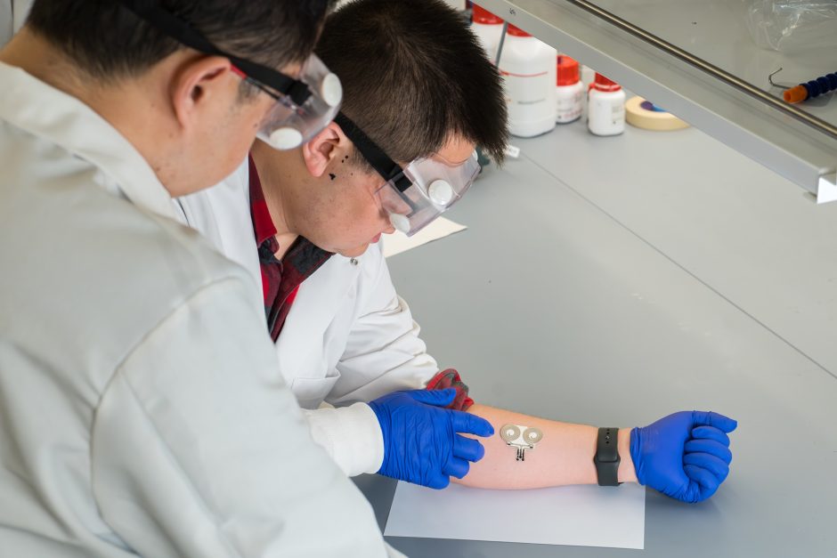
On-Skin Wearable Bioelectronic Device Paves Way for Intelligent Implants
A team of researchers at the University of Missouri (Columbia, MO, USA) has achieved a milestone in developing a state-of-the-art on-skin wearable bioelectronic device. This development comes from a lab... Read more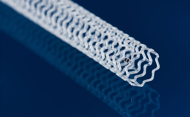
First-Of-Its-Kind Dissolvable Stent to Improve Outcomes for Patients with Severe PAD
Peripheral artery disease (PAD) affects millions and presents serious health risks, particularly its severe form, chronic limb-threatening ischemia (CLTI). CLTI develops when arteries are blocked by plaque,... Read moreSurgical Techniques
view channelHandheld Device for Fluorescence-Guided Surgery a Game Changer for Removal of High-Grade Glioma Brain Tumors
Grade III or IV gliomas are among the most common and deadly brain tumors, with around 20,000 cases annually in the U.S. and 1.2 million globally. These tumors are very aggressive and tend to infiltrate... Read more.jpg)
Cutting-Edge Robotic Bronchial Endoscopic System Provides Prompt Intervention during Emergencies
A novel robotic bronchial endoscopic system has been developed to minimize side effects and provide timely intervention for airway obstructions caused by food or foreign bodies in infants, young children,... Read morePatient Care
view channelFirst-Of-Its-Kind Portable Germicidal Light Technology Disinfects High-Touch Clinical Surfaces in Seconds
Reducing healthcare-acquired infections (HAIs) remains a pressing issue within global healthcare systems. In the United States alone, 1.7 million patients contract HAIs annually, leading to approximately... Read more
Surgical Capacity Optimization Solution Helps Hospitals Boost OR Utilization
An innovative solution has the capability to transform surgical capacity utilization by targeting the root cause of surgical block time inefficiencies. Fujitsu Limited’s (Tokyo, Japan) Surgical Capacity... Read more
Game-Changing Innovation in Surgical Instrument Sterilization Significantly Improves OR Throughput
A groundbreaking innovation enables hospitals to significantly improve instrument processing time and throughput in operating rooms (ORs) and sterile processing departments. Turbett Surgical, Inc.... Read moreHealth IT
view channel
Machine Learning Model Improves Mortality Risk Prediction for Cardiac Surgery Patients
Machine learning algorithms have been deployed to create predictive models in various medical fields, with some demonstrating improved outcomes compared to their standard-of-care counterparts.... Read more
Strategic Collaboration to Develop and Integrate Generative AI into Healthcare
Top industry experts have underscored the immediate requirement for healthcare systems and hospitals to respond to severe cost and margin pressures. Close to half of U.S. hospitals ended 2022 in the red... Read more
AI-Enabled Operating Rooms Solution Helps Hospitals Maximize Utilization and Unlock Capacity
For healthcare organizations, optimizing operating room (OR) utilization during prime time hours is a complex challenge. Surgeons and clinics face difficulties in finding available slots for booking cases,... Read more
AI Predicts Pancreatic Cancer Three Years before Diagnosis from Patients’ Medical Records
Screening for common cancers like breast, cervix, and prostate cancer relies on relatively simple and highly effective techniques, such as mammograms, Pap smears, and blood tests. These methods have revolutionized... Read morePoint of Care
view channel
Critical Bleeding Management System to Help Hospitals Further Standardize Viscoelastic Testing
Surgical procedures are often accompanied by significant blood loss and the subsequent high likelihood of the need for allogeneic blood transfusions. These transfusions, while critical, are linked to various... Read more
Point of Care HIV Test Enables Early Infection Diagnosis for Infants
Early diagnosis and initiation of treatment are crucial for the survival of infants infected with HIV (human immunodeficiency virus). Without treatment, approximately 50% of infants who acquire HIV during... Read more
Whole Blood Rapid Test Aids Assessment of Concussion at Patient's Bedside
In the United States annually, approximately five million individuals seek emergency department care for traumatic brain injuries (TBIs), yet over half of those suspecting a concussion may never get it checked.... Read more
New Generation Glucose Hospital Meter System Ensures Accurate, Interference-Free and Safe Use
A new generation glucose hospital meter system now comes with several features that make hospital glucose testing easier and more secure while continuing to offer accuracy, freedom from interference, and... Read moreBusiness
view channel
Johnson & Johnson Acquires Cardiovascular Medical Device Company Shockwave Medical
Johnson & Johnson (New Brunswick, N.J., USA) and Shockwave Medical (Santa Clara, CA, USA) have entered into a definitive agreement under which Johnson & Johnson will acquire all of Shockwave’s... Read more


