


Axillary Vein Approach Superior for ICD Leads Placement
|
By HospiMedica International staff writers Posted on 21 Jul 2020 |
|
Ultrasound guided axillary vein puncture (USAX) bests cephalic vein (CV) dissection via cut-down for pacemaker and defibrillator lead insertion, according to a new study.
Researchers at Universidade Católica do Rio Grande do Sul (PUCRS; Porto Alegre, Brazil), and Hospital de Clínicas de Porto Alegre (HCPA; Porto Alegre, Brazil) conducted a prospective, multicenter trial that included 88 adults (median age 70.5 years, 60.2% male), randomized on a 1:1 basis to either USAX or CV. The primary endpoint was defined as success rate, with secondary endpoints including venous access site change, time to obtain venous access, total procedural time, and early complication rate.
The results revealed that for the primary outcome, a higher success rate was observed in the USAX group (97.7%) than in the CV group (54.5%), as well as a lower rate of venous access site change (2.3% versus 40.9%), shorter time to obtain venous access (five minutes versus 15 minutes), and procedural time (40 minutes versus 51 minutes), with no statistical difference found in the overall complication rate (2.3% vs 11.4%). The study was published on April 29, 2020, in Heart Rhythm.
“Ultrasound-guided axillary vein puncture for implantation of pacemaker or defibrillator leads is a good first-line or alternative approach when, for example, the cephalic vein is absent or unsuitable for insertion of multiple leads,” said lead author Ana Paula Tagliari, MD, MSc, of HCPA. “Given its short procedure time, the axillary vein approach may allow a faster patient turnover. And the axillary vein itself has several advantages over the cephalic vein, including extrathoracic location, larger caliber, and greater distance from an artery.”
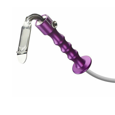
Both pacemakers and implantable cardiac devices (ICDs) have a pulse generator that fits below the collarbone, secured in a pocket under the skin, but above the muscle. The pulse generator sends pulses to send to the heart muscles via electrical leads that run through a selected large vein. The tip of a lead is anchored to a specific section of heart muscle; the rest of the lead floats freely (at least at first). Some devices have one lead, most have two, and a small but growing number have three.
Related Links:
Universidade Católica do Rio Grande do Sul
Hospital de Clínicas de Porto Alegre


Researchers at Universidade Católica do Rio Grande do Sul (PUCRS; Porto Alegre, Brazil), and Hospital de Clínicas de Porto Alegre (HCPA; Porto Alegre, Brazil) conducted a prospective, multicenter trial that included 88 adults (median age 70.5 years, 60.2% male), randomized on a 1:1 basis to either USAX or CV. The primary endpoint was defined as success rate, with secondary endpoints including venous access site change, time to obtain venous access, total procedural time, and early complication rate.
The results revealed that for the primary outcome, a higher success rate was observed in the USAX group (97.7%) than in the CV group (54.5%), as well as a lower rate of venous access site change (2.3% versus 40.9%), shorter time to obtain venous access (five minutes versus 15 minutes), and procedural time (40 minutes versus 51 minutes), with no statistical difference found in the overall complication rate (2.3% vs 11.4%). The study was published on April 29, 2020, in Heart Rhythm.
“Ultrasound-guided axillary vein puncture for implantation of pacemaker or defibrillator leads is a good first-line or alternative approach when, for example, the cephalic vein is absent or unsuitable for insertion of multiple leads,” said lead author Ana Paula Tagliari, MD, MSc, of HCPA. “Given its short procedure time, the axillary vein approach may allow a faster patient turnover. And the axillary vein itself has several advantages over the cephalic vein, including extrathoracic location, larger caliber, and greater distance from an artery.”
Both pacemakers and implantable cardiac devices (ICDs) have a pulse generator that fits below the collarbone, secured in a pocket under the skin, but above the muscle. The pulse generator sends pulses to send to the heart muscles via electrical leads that run through a selected large vein. The tip of a lead is anchored to a specific section of heart muscle; the rest of the lead floats freely (at least at first). Some devices have one lead, most have two, and a small but growing number have three.
Related Links:
Universidade Católica do Rio Grande do Sul
Hospital de Clínicas de Porto Alegre
Gold Member
POC Blood Gas Analyzer
Stat Profile Prime Plus
Gold Member
Solid State Kv/Dose Multi-Sensor
AGMS-DM+
Silver Member
Compact 14-Day Uninterrupted Holter ECG
NR-314P
New
Illuminated Retractor System
HandLite
Latest Surgical Techniques News
- Miniaturized Implantable Multi-Sensors Device to Monitor Vessels Health
- Tiny Robots Made Out Of Carbon Could Conduct Colonoscopy, Pelvic Exam or Blood Test
- Miniaturized Ultrasonic Scalpel Enables Faster and Safer Robotic-Assisted Surgery
- AI Assisted Reading Tool for Small Bowel Video Capsule Endoscopy Detects More Lesions
- First-Ever Contact Force Pulsed Field Ablation System to Transform Treatment of Ventricular Arrhythmias
- Caterpillar Robot with Built-In Steering System Crawls Easily Through Loops and Bends
- Tiny Wraparound Electronic Implants to Revolutionize Treatment of Spinal Cord Injuries
- Small, Implantable Cardiac Pump to Help Children Awaiting Heart Transplant
- Gastrointestinal Imaging Capsule a Game-Changer in Esophagus Surveillance and Treatment
- World’s Smallest Laser Probe for Brain Procedures Facilitates Ablation of Full Range of Targets
- Artificial Intelligence Broadens Diagnostic Abilities of Conventional Coronary Angiography
- AI-Powered Surgical Visualization Tool Supports Surgeons' Visual Recognition in Real Time
- Cutting-Edge Robotic Bronchial Endoscopic System Provides Prompt Intervention during Emergencies
- Handheld Device for Fluorescence-Guided Surgery a Game Changer for Removal of High-Grade Glioma Brain Tumors
- Porous Gel Sponge Facilitates Rapid Hemostasis and Wound Healing
- Novel Rigid Endoscope System Enables Deep Tissue Imaging During Surgery
Channels
Artificial Intelligence
view channel")
AI-Powered Algorithm to Revolutionize Detection of Atrial Fibrillation
Atrial fibrillation (AFib), a condition characterized by an irregular and often rapid heart rate, is linked to increased risks of stroke and heart failure. This is because the irregular heartbeat in AFib... Read more")
AI Diagnostic Tool Accurately Detects Valvular Disorders Often Missed by Doctors
Doctors generally use stethoscopes to listen for the characteristic lub-dub sounds made by heart valves opening and closing. They also listen for less prominent sounds that indicate problems with these valves.... Read more")
Critical Care
view channel")
Powerful AI Risk Assessment Tool Predicts Outcomes in Heart Failure Patients
Heart failure is a serious condition where the heart cannot pump sufficient blood to meet the body's needs, leading to symptoms like fatigue, weakness, and swelling in the legs and feet, and it can ultimately... Read more")
Peptide-Based Hydrogels Repair Damaged Organs and Tissues On-The-Spot
Scientists have ingeniously combined biomedical expertise with nature-inspired engineering to develop a jelly-like material that holds significant promise for immediate repairs to a wide variety of damaged... Read more")
One-Hour Endoscopic Procedure Could Eliminate Need for Insulin for Type 2 Diabetes
Over 37 million Americans are diagnosed with diabetes, and more than 90% of these cases are Type 2 diabetes. This form of diabetes is most commonly seen in individuals over 45, though an increasing number... Read morePatient Care
view channelFirst-Of-Its-Kind Portable Germicidal Light Technology Disinfects High-Touch Clinical Surfaces in Seconds
Reducing healthcare-acquired infections (HAIs) remains a pressing issue within global healthcare systems. In the United States alone, 1.7 million patients contract HAIs annually, leading to approximately... Read more")
Surgical Capacity Optimization Solution Helps Hospitals Boost OR Utilization
An innovative solution has the capability to transform surgical capacity utilization by targeting the root cause of surgical block time inefficiencies. Fujitsu Limited’s (Tokyo, Japan) Surgical Capacity... Read more")
Game-Changing Innovation in Surgical Instrument Sterilization Significantly Improves OR Throughput
A groundbreaking innovation enables hospitals to significantly improve instrument processing time and throughput in operating rooms (ORs) and sterile processing departments. Turbett Surgical, Inc.... Read more")
Health IT
view channel")
Machine Learning Model Improves Mortality Risk Prediction for Cardiac Surgery Patients
Machine learning algorithms have been deployed to create predictive models in various medical fields, with some demonstrating improved outcomes compared to their standard-of-care counterparts.... Read more")
Strategic Collaboration to Develop and Integrate Generative AI into Healthcare
Top industry experts have underscored the immediate requirement for healthcare systems and hospitals to respond to severe cost and margin pressures. Close to half of U.S. hospitals ended 2022 in the red... Read more")
AI-Enabled Operating Rooms Solution Helps Hospitals Maximize Utilization and Unlock Capacity
For healthcare organizations, optimizing operating room (OR) utilization during prime time hours is a complex challenge. Surgeons and clinics face difficulties in finding available slots for booking cases,... Read more")
AI Predicts Pancreatic Cancer Three Years before Diagnosis from Patients’ Medical Records
Screening for common cancers like breast, cervix, and prostate cancer relies on relatively simple and highly effective techniques, such as mammograms, Pap smears, and blood tests. These methods have revolutionized... Read morePoint of Care
view channel clearance for use with its Quantra QStat Cartridge (Photo courtesy of HemoSonics)")
Critical Bleeding Management System to Help Hospitals Further Standardize Viscoelastic Testing
Surgical procedures are often accompanied by significant blood loss and the subsequent high likelihood of the need for allogeneic blood transfusions. These transfusions, while critical, are linked to various... Read more prequalification (Photo courtesy of Cepheid)")
Point of Care HIV Test Enables Early Infection Diagnosis for Infants
Early diagnosis and initiation of treatment are crucial for the survival of infants infected with HIV (human immunodeficiency virus). Without treatment, approximately 50% of infants who acquire HIV during... Read more")
Whole Blood Rapid Test Aids Assessment of Concussion at Patient's Bedside
In the United States annually, approximately five million individuals seek emergency department care for traumatic brain injuries (TBIs), yet over half of those suspecting a concussion may never get it checked.... Read more")
New Generation Glucose Hospital Meter System Ensures Accurate, Interference-Free and Safe Use
A new generation glucose hospital meter system now comes with several features that make hospital glucose testing easier and more secure while continuing to offer accuracy, freedom from interference, and... Read moreBusiness
view channel")
Johnson & Johnson Acquires Cardiovascular Medical Device Company Shockwave Medical
Johnson & Johnson (New Brunswick, N.J., USA) and Shockwave Medical (Santa Clara, CA, USA) have entered into a definitive agreement under which Johnson & Johnson will acquire all of Shockwave’s... Read more")
")