Point-of-Care Ultrasound Predicts Clinical Outcomes in Patients with COVID-19
|
By HospiMedica International staff writers Posted on 06 Sep 2021 |
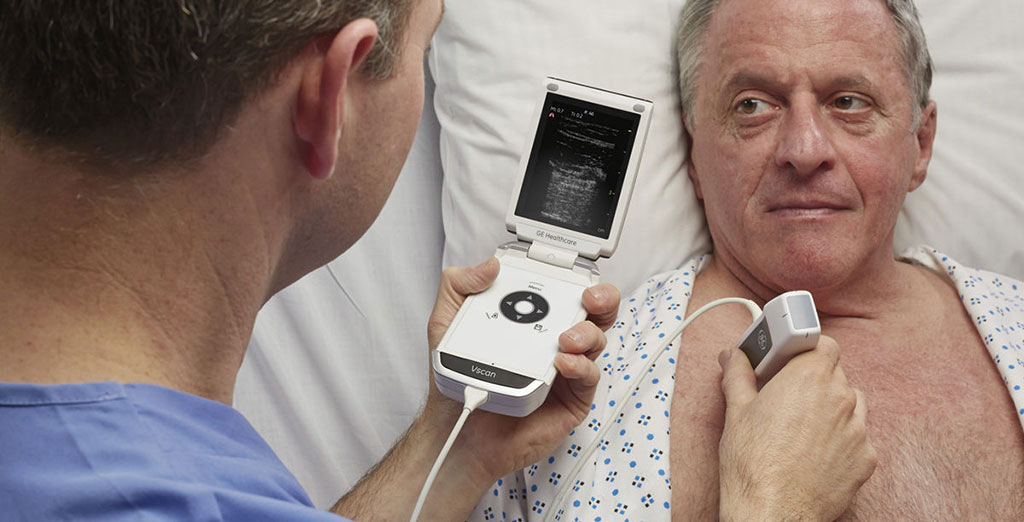
Image: GE Healthcare Vscan (Photo courtesy of GE Healthcare)
Point-of-care ultrasound (POCUS) can detect the pulmonary manifestations of COVID-19 and predict patient outcomes, according to a new study.
These findings were from a prospective cohort study conducted by researchers of Stanford University (Stanford, CA, USA) at four hospitals from March 2020 to January 2021 to evaluate lung POCUS and clinical outcomes of COVID-19. Inclusion criteria included adult patients hospitalized for COVID-19 who received lung POCUS with a 12-zone protocol. Each image was interpreted by two reviewers blinded to clinical outcomes. The study’s primary outcome was the need for intensive care unit (ICU) admission versus no ICU admission. Secondary outcomes included intubation and supplemental oxygen usage.
POCUS has garnered substantial interest as a potential modality to expediently diagnose COVID-19 and its complications. POCUS devices are cheaper than traditional imaging equipment, such as X-ray or computed tomography (CT) machines, which makes POCUS ideal for surge scenarios and resource-limited settings. Since providers using POCUS are concomitantly at the bedside assessing patients, POCUS permits an immediate and augmented evaluation of the patient. It can reduce personal protective equipment usage by radiology technicians as well as the need to decontaminate larger radiographic equipment. POCUS has also been successfully used in the diagnosis and management of COVID-19. Previously described pulmonary manifestations of COVID-19 include pulmonary edema, lung consolidation, and pleural-line irregularities. POCUS can diagnose these pathological states with similar accuracy to CT and with higher sensitivity than X-ray.
Although lung ultrasound abnormalities are more common in patients who experience adverse outcomes with COVID-19, few studies have examined whether scans performed early in the hospitalization can provide meaningful risk stratification. Furthermore, few scoring tools predict the need for oxygen on discharge, which represents a limited resource in many settings. In the latest study, the researchers examined whether early pulmonary POCUS findings correlate with important clinical outcomes, such as intensive care admission or need for supplemental oxygen. They also examined whether these findings, if detected early, are predictive of future clinical outcomes in the subsequent hospital course or after discharge.
In this prospective cohort study conducted at four medical centers of patients hospitalized with COVID-19, the researchers found that lung ultrasounds collected within 24 hours of emergency department triage were predictive of important clinical outcomes in the subsequent hospital course, including ICU admission, intubation, supplemental oxygen usage, and the need for oxygen at discharge. Ultrasound findings associated with an adverse clinical course included B-lines and consolidations (particularly in the anterior and lateral lung fields), while a normal ultrasound on triage was protective against adverse outcomes. Notably, ultrasound findings did not dynamically change over a 28-day window after symptom onset, suggesting that the presence of B-lines or consolidations, regardless of when they are detected, may be important clinical predictors.
Previous investigations have demonstrated that lung POCUS findings (such as B-lines or consolidations) are associated with critical illness and intubation for COVID-19. The new study expands on these observations by demonstrating that scans collected within 24 hours of ED triage may predict outcomes for the entire hospital course, including future supplemental oxygen usage and the need for oxygen on discharge. This information may substantially aid frontline providers in resource-limited settings experiencing patient surges. In such scenarios, POCUS could augment admission or discharge decisions for providers. More broadly, POCUS could represent one of several tools to identify patients at-risk for adverse outcomes. Other authors have demonstrated the utility of laboratory tests (eg, ferritin, c-reactive protein) or radiographic findings for risk stratification. POCUS may have potential advantages over these other methods in that it is more expedient, low cost and does not expose the patient to ionizing radiation. Future studies are needed to directly compare POCUS with other scoring systems that utilize laboratory or radiological findings.
Related Links:
Stanford University
Latest COVID-19 News
- Low-Cost System Detects SARS-CoV-2 Virus in Hospital Air Using High-Tech Bubbles
- World's First Inhalable COVID-19 Vaccine Approved in China
- COVID-19 Vaccine Patch Fights SARS-CoV-2 Variants Better than Needles
- Blood Viscosity Testing Can Predict Risk of Death in Hospitalized COVID-19 Patients
- ‘Covid Computer’ Uses AI to Detect COVID-19 from Chest CT Scans
- MRI Lung-Imaging Technique Shows Cause of Long-COVID Symptoms
- Chest CT Scans of COVID-19 Patients Could Help Distinguish Between SARS-CoV-2 Variants
- Specialized MRI Detects Lung Abnormalities in Non-Hospitalized Long COVID Patients
- AI Algorithm Identifies Hospitalized Patients at Highest Risk of Dying From COVID-19
- Sweat Sensor Detects Key Biomarkers That Provide Early Warning of COVID-19 and Flu
- Study Assesses Impact of COVID-19 on Ventilation/Perfusion Scintigraphy
- CT Imaging Study Finds Vaccination Reduces Risk of COVID-19 Associated Pulmonary Embolism
- Third Day in Hospital a ‘Tipping Point’ in Severity of COVID-19 Pneumonia
- Longer Interval Between COVID-19 Vaccines Generates Up to Nine Times as Many Antibodies
- AI Model for Monitoring COVID-19 Predicts Mortality Within First 30 Days of Admission
- AI Predicts COVID Prognosis at Near-Expert Level Based Off CT Scans
Channels
Critical Care
view channel
Mechanosensing-Based Approach Offers Promising Strategy to Treat Cardiovascular Fibrosis
Cardiac fibrosis, which involves the stiffening and scarring of heart tissue, is a fundamental feature of nearly every type of heart disease, from acute ischemic injuries to genetic cardiomyopathies.... Read more
AI Interpretability Tool for Photographed ECG Images Offers Pixel-Level Precision
The electrocardiogram (ECG) is a crucial diagnostic tool in modern medicine, used to detect heart conditions such as arrhythmias and structural abnormalities. Every year, millions of ECGs are performed... Read moreSurgical Techniques
view channel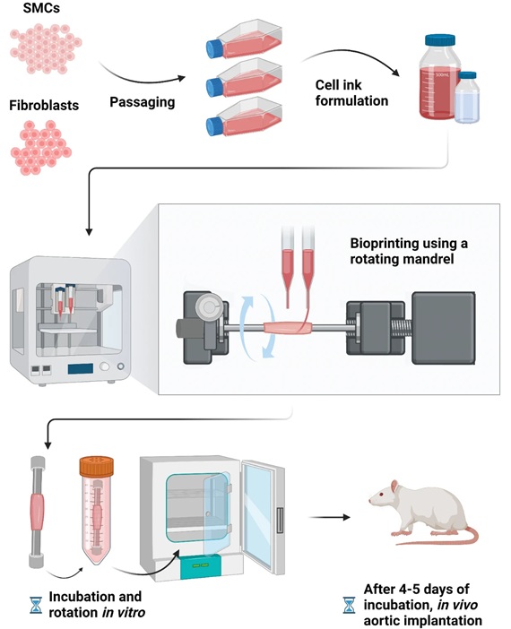
Bioprinted Aortas Offer New Hope for Vascular Repair
Current treatment options for severe cardiovascular diseases include using grafts made from a patient's own tissue (autologous) or synthetic materials. However, autologous grafts require invasive surgery... Read more
Early TAVR Intervention Reduces Cardiovascular Events in Asymptomatic Aortic Stenosis Patients
Each year, approximately 300,000 Americans are diagnosed with aortic stenosis (AS), a serious condition that results from the narrowing or blockage of the aortic valve in the heart. Two common treatments... Read more
New Procedure Found Safe and Effective for Patients Undergoing Transcatheter Mitral Valve Replacement
In the United States, approximately four million people suffer from mitral valve regurgitation, the most common type of heart valve disease. As an alternative to open-heart surgery, transcatheter mitral... Read morePatient Care
view channel
Portable Biosensor Platform to Reduce Hospital-Acquired Infections
Approximately 4 million patients in the European Union acquire healthcare-associated infections (HAIs) or nosocomial infections each year, with around 37,000 deaths directly resulting from these infections,... Read moreFirst-Of-Its-Kind Portable Germicidal Light Technology Disinfects High-Touch Clinical Surfaces in Seconds
Reducing healthcare-acquired infections (HAIs) remains a pressing issue within global healthcare systems. In the United States alone, 1.7 million patients contract HAIs annually, leading to approximately... Read more
Surgical Capacity Optimization Solution Helps Hospitals Boost OR Utilization
An innovative solution has the capability to transform surgical capacity utilization by targeting the root cause of surgical block time inefficiencies. Fujitsu Limited’s (Tokyo, Japan) Surgical Capacity... Read more
Game-Changing Innovation in Surgical Instrument Sterilization Significantly Improves OR Throughput
A groundbreaking innovation enables hospitals to significantly improve instrument processing time and throughput in operating rooms (ORs) and sterile processing departments. Turbett Surgical, Inc.... Read moreHealth IT
view channel
Printable Molecule-Selective Nanoparticles Enable Mass Production of Wearable Biosensors
The future of medicine is likely to focus on the personalization of healthcare—understanding exactly what an individual requires and delivering the appropriate combination of nutrients, metabolites, and... Read more
Smartwatches Could Detect Congestive Heart Failure
Diagnosing congestive heart failure (CHF) typically requires expensive and time-consuming imaging techniques like echocardiography, also known as cardiac ultrasound. Previously, detecting CHF by analyzing... Read moreBusiness
view channel
Expanded Collaboration to Transform OR Technology Through AI and Automation
The expansion of an existing collaboration between three leading companies aims to develop artificial intelligence (AI)-driven solutions for smart operating rooms with sophisticated monitoring and automation.... Read more


