


Magnetic Actuation Device Enhances Laparoscopic Surgery 
|
By HospiMedica International staff writers Posted on 15 Mar 2015 |
|
.")
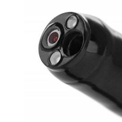
Image: An early prototype of the internal retractor unit (Photo courtesy of Vanderbilt University).
Two new studies describe a local magnetic actuation (LMA) approach using coaxial gears to ease tissue manipulation during minimally invasive surgery (MIS) procedures.
Researchers at Vanderbilt University (Nashville, TN, USA) developed An LMA actuation unit that consists of a pair of diametrically magnetized single-dipole cylindrical magnets, working as a gear system that crosses the abdominal wall. In principal, one unit is external, placed on a patient’s abdomen; the second is an internal unit small enough to fit through the access ports used in MIS. During operation, the internal unit is magnetically anchored to the inside of the abdominal wall, while the other unit provides the mechanical force that powers the device.
The magnet in the external unit is attached to the shaft of a powerful electric motor that causes it to spin. The magnet in the internal unit is also attached to a shaft, but one that drives a two-inch lever. When the electric motor on the external unit twirls its magnet, it generates a rotating magnetic field that forces the magnet on the shaft in the inner unit to spin at the same speed. When it spins in one direction, the lever opens up, and when it spins in the opposite direction, the lever closes.
To retract an organ, the surgeon inserts the internal unit with a laparoscopic grasper and inserts it through the port into the body. When the internal unit is close enough to the external unit, it snaps into position against the inner surface of the abdominal wall. The motor on the external unit is then engaged, lowering the lever. Using standard laparoscopic instruments, the surgeon attaches one end of a line to the tip of the lever and the other end to a clip or suction cup fastened to the organ that must be moved. The electric motor is run in reverse and the lever retracts, pulling the organ into the desired position.
The first LMA device the researchers built as proof of principle was used during liver resection in vivo on an anesthetized porcine model. The researchers found that when abdominal wall thickness is 2 cm, the retractor is able to lift more than ten times its own weight. The researchers concluded that LMA can enable the transfer of a larger amount of mechanical power than that possible by using motors on the laparoscopic instrument itself. The studies were published in the February 2015 issue of IEEE Transactions on Robotics and the March 2015 issue of ASME Journal of Medical Devices.
“This device demonstrates for the first time that controllable mechanical power can be transferred across the abdominal wall via an intelligent magnetic link to power a robotic instrument,” said lead author Assistant Professor of Mechanical Engineering Pietro Valdastri, MSc, PhD. “Besides the ability to deliver a lot of power, the magnetic actuation approach has some other important advantages; the internal units do not contain any expensive and delicate electronics, so they can be easily sterilized and, if manufactured in bulk, could be made inexpensively enough to be disposable.”
Related Links:
Vanderbilt University
Gold Member
Real-Time Diagnostics Onscreen Viewer
GEMweb Live
Gold Member
Disposable Protective Suit For Medical Use
Disposable Protective Suit For Medical Use
Silver Member
Compact 14-Day Uninterrupted Holter ECG
NR-314P
New
Bronchoscope
EB-500
Latest Surgical Techniques News
- Flexible Microdisplay Visualizes Brain Activity in Real-Time To Guide Neurosurgeons
- Next-Gen Computer Assisted Vacuum Thrombectomy Technology Rapidly Removes Blood Clots
- Hydrogel-Based Miniaturized Electric Generators to Power Biomedical Devices
- Custom 3D-Printed Orthopedic Implants Transform Joint Replacement Surgery
- Wearable Technology Monitors and Analyzes Surgeons' Posture during Long Surgical Procedures
- Cutting-Edge Imaging Platform Detects Residual Breast Cancer Missed During Lumpectomy Surgery
- Computational Models Predict Heart Valve Leakage in Children
- Breakthrough Device Enables Clear and Real-Time Visual Guidance for Effective Cardiovascular Interventions
- World’s First Microscopic Probe to Revolutionize Early Cancer Diagnosis
- World’s Smallest Implantable Brain Stimulator Demonstrated in Human Patient
- Robotically Assisted Lung Transplants Could Soon Become a Reality
- AI to Provide Heart Transplant Surgeons with New Decision-Making Data
- New Surgical Tool Empowers Precision and Confidence in Operating Room
- Future Muscle-Powered Surgical Robots Could Perform Minimally Invasive Procedures inside Body
- Non-Invasive Technique Combines Cardiac CT with AI-Powered Blood Flow for Heart Bypass Surgery
- First-Of-Its-Kind Device Repairs Leaky Tricuspid Heart Valve
Channels
Artificial Intelligence
view channel")
AI-Powered Algorithm to Revolutionize Detection of Atrial Fibrillation
Atrial fibrillation (AFib), a condition characterized by an irregular and often rapid heart rate, is linked to increased risks of stroke and heart failure. This is because the irregular heartbeat in AFib... Read more")
AI Diagnostic Tool Accurately Detects Valvular Disorders Often Missed by Doctors
Doctors generally use stethoscopes to listen for the characteristic lub-dub sounds made by heart valves opening and closing. They also listen for less prominent sounds that indicate problems with these valves.... Read more")
")
Critical Care
view channel")
Wheeze-Counting Wearable Device Monitors Patient's Breathing In Real Time
Lung diseases like asthma, chronic obstructive pulmonary disease (COPD), lung cancer, bronchitis, and infections such as pneumonia, rank among the leading causes of death worldwide. Traditionally, medical... Read more")
Wearable Multiplex Biosensors Could Revolutionize COPD Management
Chronic obstructive pulmonary disease (COPD) ranks as the third leading cause of death worldwide. Acute exacerbations of COPD (AECOPD), which are often triggered by lung infections, accelerate the disease's... Read more.jpg "Image: New machine learning models can help solve the problem of underdiagnosed heart disease in women (Photo courtesy of 123RF)")
Patient Care
view channel")
Surgical Capacity Optimization Solution Helps Hospitals Boost OR Utilization
An innovative solution has the capability to transform surgical capacity utilization by targeting the root cause of surgical block time inefficiencies. Fujitsu Limited’s (Tokyo, Japan) Surgical Capacity... Read more")
Game-Changing Innovation in Surgical Instrument Sterilization Significantly Improves OR Throughput
A groundbreaking innovation enables hospitals to significantly improve instrument processing time and throughput in operating rooms (ORs) and sterile processing departments. Turbett Surgical, Inc.... Read more")
Next Gen ICU Bed to Help Address Complex Critical Care Needs
As the critical care environment becomes increasingly demanding and complex due to evolving hospital needs, there is a pressing requirement for innovations that can facilitate patient recovery.... Read moreGroundbreaking AI-Powered UV-C Disinfection Technology Redefines Infection Control Landscape
Healthcare-associated infection (HCAI) is a widespread complication in healthcare management, posing a significant health risk due to its potential to increase patient morbidity and mortality, prolong... Read moreHealth IT
view channel")
Machine Learning Model Improves Mortality Risk Prediction for Cardiac Surgery Patients
Machine learning algorithms have been deployed to create predictive models in various medical fields, with some demonstrating improved outcomes compared to their standard-of-care counterparts.... Read more")
Strategic Collaboration to Develop and Integrate Generative AI into Healthcare
Top industry experts have underscored the immediate requirement for healthcare systems and hospitals to respond to severe cost and margin pressures. Close to half of U.S. hospitals ended 2022 in the red... Read more")
AI-Enabled Operating Rooms Solution Helps Hospitals Maximize Utilization and Unlock Capacity
For healthcare organizations, optimizing operating room (OR) utilization during prime time hours is a complex challenge. Surgeons and clinics face difficulties in finding available slots for booking cases,... Read more")
AI Predicts Pancreatic Cancer Three Years before Diagnosis from Patients’ Medical Records
Screening for common cancers like breast, cervix, and prostate cancer relies on relatively simple and highly effective techniques, such as mammograms, Pap smears, and blood tests. These methods have revolutionized... Read morePoint of Care
view channel clearance for use with its Quantra QStat Cartridge (Photo courtesy of HemoSonics)")
Critical Bleeding Management System to Help Hospitals Further Standardize Viscoelastic Testing
Surgical procedures are often accompanied by significant blood loss and the subsequent high likelihood of the need for allogeneic blood transfusions. These transfusions, while critical, are linked to various... Read more prequalification (Photo courtesy of Cepheid)")
Point of Care HIV Test Enables Early Infection Diagnosis for Infants
Early diagnosis and initiation of treatment are crucial for the survival of infants infected with HIV (human immunodeficiency virus). Without treatment, approximately 50% of infants who acquire HIV during... Read more")
Whole Blood Rapid Test Aids Assessment of Concussion at Patient's Bedside
In the United States annually, approximately five million individuals seek emergency department care for traumatic brain injuries (TBIs), yet over half of those suspecting a concussion may never get it checked.... Read more")
New Generation Glucose Hospital Meter System Ensures Accurate, Interference-Free and Safe Use
A new generation glucose hospital meter system now comes with several features that make hospital glucose testing easier and more secure while continuing to offer accuracy, freedom from interference, and... Read moreBusiness
view channel")
Johnson & Johnson Acquires Cardiovascular Medical Device Company Shockwave Medical
Johnson & Johnson (New Brunswick, N.J., USA) and Shockwave Medical (Santa Clara, CA, USA) have entered into a definitive agreement under which Johnson & Johnson will acquire all of Shockwave’s... Read more")
")