Computer Vision Analyzes Stroke Rehabilitation Process
|
By HospiMedica International staff writers Posted on 31 Jan 2018 |
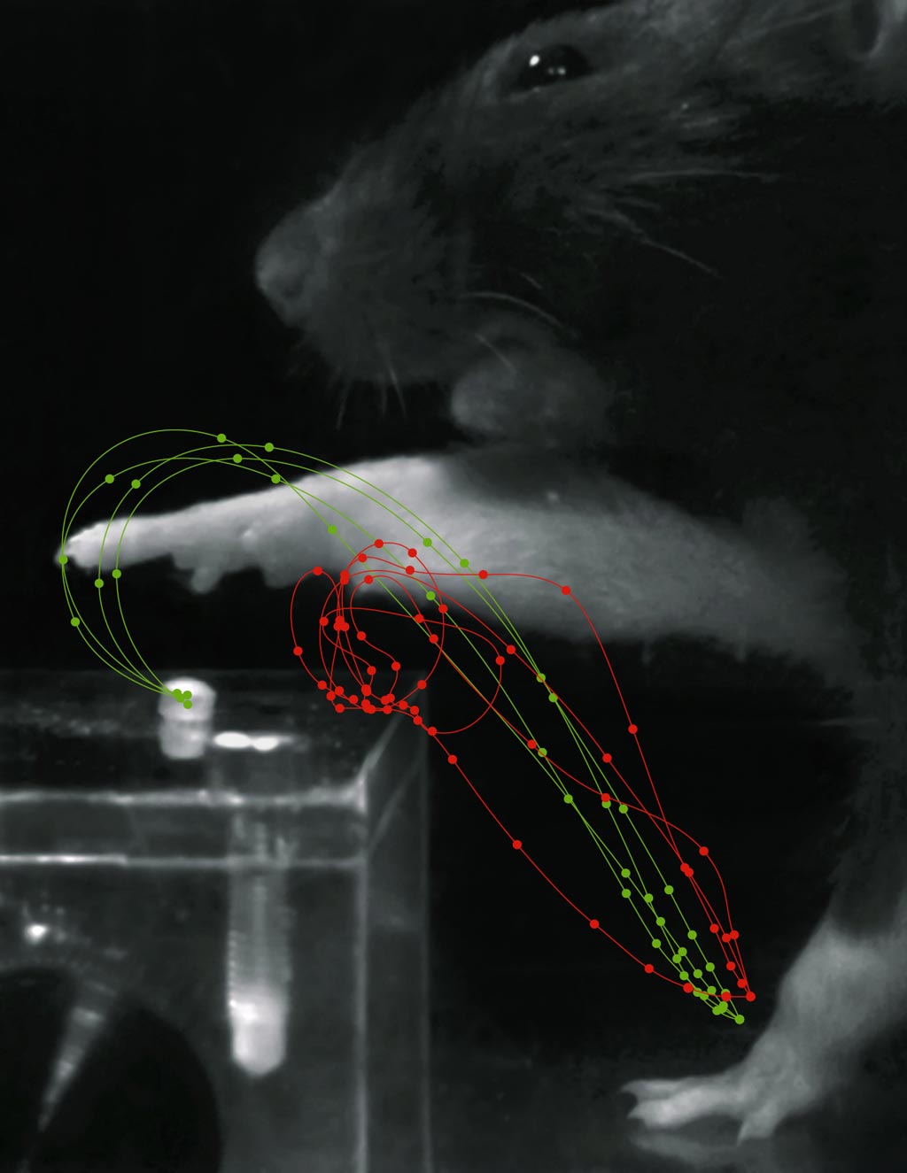
Image: Red trajectories show grasp movements after stroke, while green trajectories show rehabilitation (Photo courtesy of Tabea Kraus/ ETH).
A new study shows how optogenetics and machine learning can be used to analyze changes in motor skills, aiding stroke patient recovery.
Developed at the Swiss Federal Institute of Technology (ETH; Zurich, Switzerland), the University of Heidelberg (Germany), and other institutions, the new therapeutic approach is based on using optogenetics to activate corticospinal circuitry. The optogenetic stimulation, in conjunction with intense, scheduled rehabilitation can lead to the restoration of lost movement patterns (rather than induced compensatory actions), as revealed by a computer vision-based automatic behavior analysis in a stroke model study in rats.
The rat movements were recorded with a video camera and automatically analyzed to monitor the rehabilitation process to adjust the optogenetic stimulation. The results revealed that optogenetically activated corticospinal neurons promote axonal sprouting from the intact to the denervated cervical hemi-cord. Conversely, silencing subsets of corticospinal neurons in the recovered animals resulted in mistargeting of restored grasping function, identifying reestablishment of specific, anatomically localized cortical microcircuits. The study was published on October 30, 2017, in Nature Communications.
“Using our automatic evaluation of the movement processes, we were able to demonstrate a full recovery,” said senior author Professor Björn Ommer, PhD, of the Heidelberg University Interdisciplinary Center for Scientific Computing (IWR). “The new computer vision technique is able to quantify even the slightest changes in motor functions. By recording and analyzing the movements, we can objectively assess whether there was true restoration of the original function or merely compensation.”
“Neurorehabilitation is the only treatment option for the majority of stroke victims. Many approaches in basic science and in the clinic aim to trigger regeneration processes post-stroke by stimulating healthy brain regions of indeterminate size,” said lead author neuroscientist Anna-Sophia Wahl, MD, PhD, of ETH. “These results provide a conceptual framework to improve established clinical techniques such as transcranial magnetic or transcranial direct current stimulation in stroke patients.”
Optogenetics is a biological technique which involves the use of light to control cells in living tissue that have been genetically modified to express light-sensitive ion channels. For neuromodulation, it is used to control and monitor the activities of individual neurons in living tissue. Key reagents in optogenetics include light-sensitive proteins, such as channelrhodopsin, halorhodopsin, and archaerhodopsin, while optical recording of neuronal activities can be made with the help of optogenetic sensors for calcium (GCaMP), vesicular release (synapto-pHluorin), Neurotransmitter (GluSnFRs), or membrane voltage.
Related Links:
Swiss Federal Institute of Technology
University of Heidelberg
Latest Patient Care News
- Revolutionary Automatic IV-Line Flushing Device to Enhance Infusion Care
- VR Training Tool Combats Contamination of Portable Medical Equipment
- Portable Biosensor Platform to Reduce Hospital-Acquired Infections
- First-Of-Its-Kind Portable Germicidal Light Technology Disinfects High-Touch Clinical Surfaces in Seconds
- Surgical Capacity Optimization Solution Helps Hospitals Boost OR Utilization

- Game-Changing Innovation in Surgical Instrument Sterilization Significantly Improves OR Throughput
- Next Gen ICU Bed to Help Address Complex Critical Care Needs
- Groundbreaking AI-Powered UV-C Disinfection Technology Redefines Infection Control Landscape
- Clean Hospitals Can Reduce Antibiotic Resistance, Save Lives
- Smart Hospital Beds Improve Accuracy of Medical Diagnosis
- New Fast Endoscope Drying System Improves Productivity and Traceability
- World’s First Automated Endoscope Cleaner Fights Antimicrobial Resistance
- Portable High-Capacity Digital Stretcher Scales Provide Precision Weighing for Patients in ER
- Portable Clinical Scale with Remote Indicator Allows for Flexible Patient Weighing Use
- Innovative and Highly Customizable Medical Carts Offer Unlimited Configuration Possibilities
- Biomolecular Wound Healing Film Adheres to Sensitive Tissue and Releases Active Ingredients
Channels
Critical Care
view channel
CPR Guidelines Updated for Pediatric and Neonatal Emergency Care and Resuscitation
Cardiac arrest in infants and children remains a leading cause of pediatric emergencies, with more than 7,000 out-of-hospital and 20,000 in-hospital cardiac arrests occurring annually in the United States.... Read more
Ingestible Capsule Monitors Intestinal Inflammation
Acute mesenteric ischemia—a life-threatening condition caused by blocked blood flow to the intestines—remains difficult to diagnose early because its symptoms often mimic common digestive problems.... Read more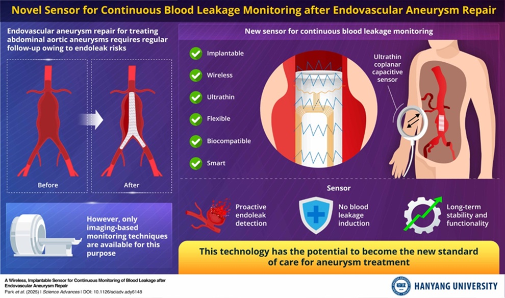
Wireless Implantable Sensor Enables Continuous Endoleak Monitoring
Endovascular aneurysm repair (EVAR) is a life-saving, minimally invasive treatment for abdominal aortic aneurysms—balloon-like bulges in the aorta that can rupture with fatal consequences.... Read more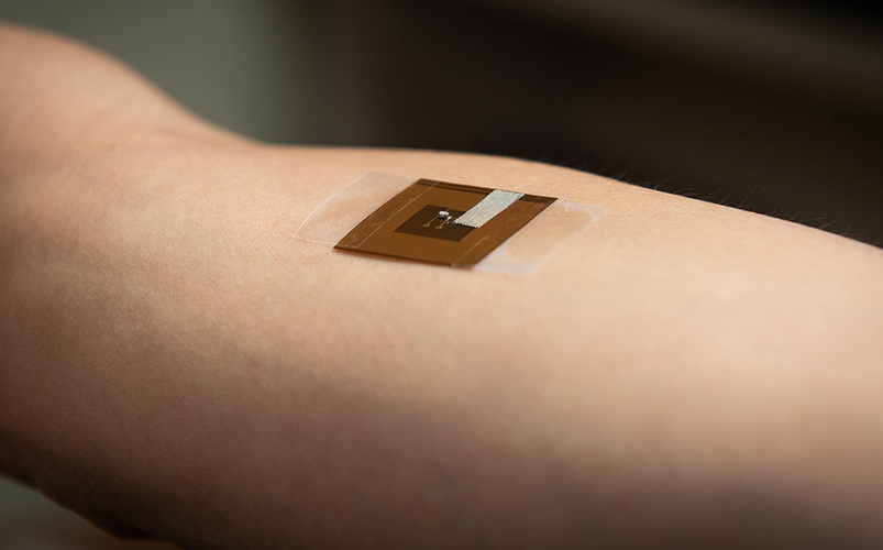
Wearable Patch for Early Skin Cancer Detection to Reduce Unnecessary Biopsies
Skin cancer remains one of the most dangerous and common cancers worldwide, with early detection crucial for improving survival rates. Traditional diagnostic methods—visual inspections, imaging, and biopsies—can... Read moreSurgical Techniques
view channel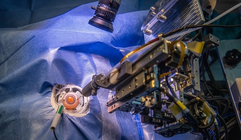
Robotic Assistant Delivers Ultra-Precision Injections with Rapid Setup Times
Age-related macular degeneration (AMD) is a leading cause of blindness worldwide, affecting nearly 200 million people, a figure expected to rise to 280 million by 2040. Current treatment involves doctors... Read more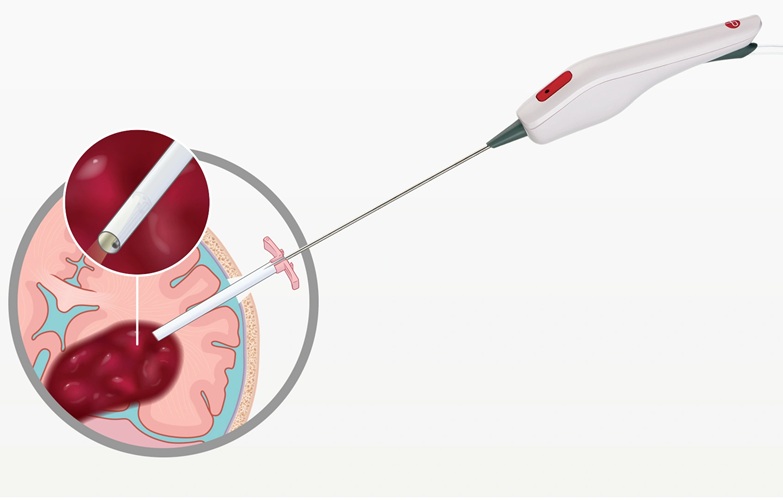
Minimally Invasive Endoscopic Surgery Improves Severe Stroke Outcomes
Intracerebral hemorrhage, a type of stroke caused by bleeding deep within the brain, remains one of the most challenging neurological emergencies to treat. Accounting for about 15% of all strokes, it carries... Read moreHealth IT
view channel
Printable Molecule-Selective Nanoparticles Enable Mass Production of Wearable Biosensors
The future of medicine is likely to focus on the personalization of healthcare—understanding exactly what an individual requires and delivering the appropriate combination of nutrients, metabolites, and... Read moreBusiness
view channel
Philips and Masimo Partner to Advance Patient Monitoring Measurement Technologies
Royal Philips (Amsterdam, Netherlands) and Masimo (Irvine, California, USA) have renewed their multi-year strategic collaboration, combining Philips’ expertise in patient monitoring with Masimo’s noninvasive... Read more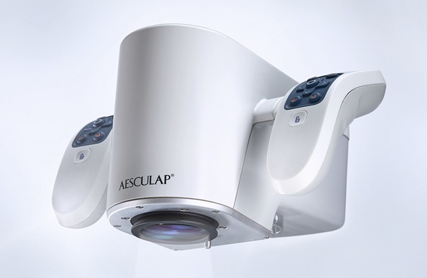
B. Braun Acquires Digital Microsurgery Company True Digital Surgery
The high-end microsurgery market in neurosurgery, spine, and ENT is undergoing a significant transformation. Traditional analog microscopes are giving way to digital exoscopes, which provide improved visualization,... Read more
CMEF 2025 to Promote Holistic and High-Quality Development of Medical and Health Industry
The 92nd China International Medical Equipment Fair (CMEF 2025) Autumn Exhibition is scheduled to be held from September 26 to 29 at the China Import and Export Fair Complex (Canton Fair Complex) in Guangzhou.... Read more