


Thermal Imaging Improves Diabetes-Related Ulcer Assessment
|
By HospiMedica International staff writers Posted on 14 Jul 2021 |
|
")
Image: Thermal images of a venous leg ulcer that ultimately failed to heal (Photo courtesy of RMIT)
A new suggests that textural analysis of thermal images of venous leg ulcers (VLUs) can detect whether a wound will need extra management.
Researchers at RMIT University (RMIT; Melbourne, Australia) and the Bolton Clarke Research Institute (Melbourne, Australia) conducted a study involving 60 patients with 72 VLUs who were followed over a period of twelve weeks. Digital and thermal images, as well as planimetry wound tracings were recorded, with the weekly change in thermal textural features computed. VLU’s were labelled as healed or unhealed based on status of the wound at 12th week follow up.
The results showed that principal component analysis (PCA) of the change in textural features in the second week (compared to week 0) were statistically significant predicting which VLA will not heal by week 12. According to the researchers, this is because wounds change significantly over their healing trajectory, with higher temperatures signaling potential inflammation or infection, while lower temperatures indicate a slower healing rate due to decreased oxygen in the region. The study was published on June 24, 2021, in the Nature Scientific Reports.
“The significance of this work is that there is now a method for detecting wounds that do not heal in the normal trajectory by week two using a non-contact, quick, objective, and simple method,” said study co-author Rajna Ogrin, PhD, of Bolton Clarke Research Institute. “A non-contact method like thermal imaging would be ideal to use when managing wounds in the home setting to minimize physical contact, and therefore reduce infection risk.”
VLU guidelines state that if wound area has not reduced by at least 20–40% after four weeks, additional interventions are warranted. Current assessment methods to monitor wound progress over this four week period involve wound tracing and measurement, including use of digital planimetry, observation and documentation of the wound tissue type, wound edge characteristics, peri-wound and surrounding skin state, and level and type of exudate. This requires at least weekly evaluations and physical contact with the wound.
Related Links:
RMIT University
Bolton Clarke Research Institute
New
Gold Member
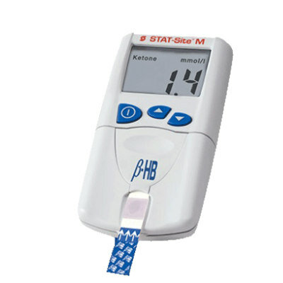
Handheld Blood Glucose Analyzer
STAT-Site
New
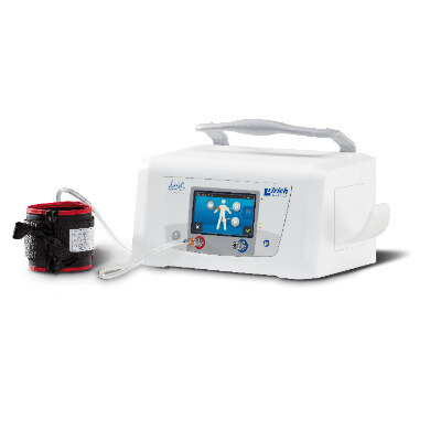
Tourniquet System
heidi– mein Tourniquet
New
Gas Analyzer
GE SAM
Latest Patient Care News
- Wearable Sleep Data Predict Adherence to Pulmonary Rehabilitation
- Revolutionary Automatic IV-Line Flushing Device to Enhance Infusion Care
- VR Training Tool Combats Contamination of Portable Medical Equipment
- Portable Biosensor Platform to Reduce Hospital-Acquired Infections
- First-Of-Its-Kind Portable Germicidal Light Technology Disinfects High-Touch Clinical Surfaces in Seconds
- Surgical Capacity Optimization Solution Helps Hospitals Boost OR Utilization

- Game-Changing Innovation in Surgical Instrument Sterilization Significantly Improves OR Throughput
- Next Gen ICU Bed to Help Address Complex Critical Care Needs
- Groundbreaking AI-Powered UV-C Disinfection Technology Redefines Infection Control Landscape
- Clean Hospitals Can Reduce Antibiotic Resistance, Save Lives
- Smart Hospital Beds Improve Accuracy of Medical Diagnosis
- New Fast Endoscope Drying System Improves Productivity and Traceability
- World’s First Automated Endoscope Cleaner Fights Antimicrobial Resistance
- Portable High-Capacity Digital Stretcher Scales Provide Precision Weighing for Patients in ER
- Portable Clinical Scale with Remote Indicator Allows for Flexible Patient Weighing Use
- Innovative and Highly Customizable Medical Carts Offer Unlimited Configuration Possibilities
Channels
Artificial Intelligence
view channelAI Analysis of Pericardial Fat Refines Long-Term Heart Disease Risk
Accurately identifying long-term cardiovascular disease risk in asymptomatic adults remains challenging for clinicians. Missed or underestimated risk delays preventive therapy and increases the chance... Read more")
Machine Learning Approach Enhances Liver Cancer Risk Stratification
Hepatocellular carcinoma, the most common form of primary liver cancer, is often detected late despite targeted surveillance programs. Current screening guidelines emphasize patients with known cirrhosis,... Read more")
, k-means++ was used to identify clusters. Three clinically distinct clusters were identified. Among those clusters, patients were divided into solid organ transplant (SOT) and non-SOT groups. Clinical characteristics and outcomes were evaluated (Masayuki Nigo et al., American Journal of Transplantation (2026). DOI: 10.1016/j.ajt.2025.10.019)")
Critical Care
view channel")
Angiography-Based FFR Approach Matches Gold Standard Results Without Wires
Accurately determining whether a coronary stenosis limits blood flow is essential to guide percutaneous coronary intervention, yet wire-based physiologic testing remains underused due to added procedural... Read more")
Eye Imaging AI Identifies Elevated Cardiovascular Risk
Many adults at risk for atherosclerotic cardiovascular disease are not identified until they undergo formal primary care assessment. Delayed risk recognition can postpone initiation of statins and lifestyle... Read more")
")
Surgical Techniques
view channel")
Fiber-Form Bone Graft Expands Intraoperative Options for Spinal Fusion
Spinal and orthopedic fusion procedures often require bone graft materials that handle predictably and support bone formation. Surgeons face added complexity in difficult anatomy and challenging fusion environments.... Read more")
Ultrasound‑Aided Catheter Treatment Cuts Early Collapse in Pulmonary Embolism
Acute pulmonary embolism can cause rapid hemodynamic deterioration and early death in hospitalized and emergency patients. Systemic thrombolysis can dissolve clots but is limited by a high risk of major... Read more")
")
Health IT
view channel")
Voice-Driven AI System Enables Structured GI Procedure Documentation
Documentation during gastrointestinal (GI) procedures often competes with real-time clinical decision-making and imposes a significant cognitive burden on physicians. Manual data entry and post-procedure... Read more")
EMR-Based Tool Predicts Graft Failure After Kidney Transplant
Kidney transplantation offers patients with end-stage kidney disease longer survival and better quality of life than dialysis, yet graft failure remains a major challenge. Although a successful transplant... Read more")
Printable Molecule-Selective Nanoparticles Enable Mass Production of Wearable Biosensors
The future of medicine is likely to focus on the personalization of healthcare—understanding exactly what an individual requires and delivering the appropriate combination of nutrients, metabolites, and... Read more")
")
Business
view channel")
")
")