New AI Research to Help Predict COVID-19 Resource Needs from Series of X-Rays
|
By HospiMedica International staff writers Posted on 18 Jan 2021 |
Illustration
Scientists have developed machine learning (ML) models that could help doctors predict how a COVID-19 patient’s condition may develop, in order to help hospitals ensure they have sufficient resources to care for patients.
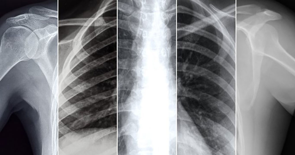
Facebook AI (Menlo Park, CA, USA), in collaboration with NYU Langone Health’s Predictive Analytics Unit and Department of Radiology (Brooklyn, NY, USA) has developed three ML models that can help hospitals know whether COVID-19 patients are likely to need escalated treatment and plan accordingly. The first ML model can predict patient deterioration based on a single X-ray and the second model can predict patient deterioration based on a sequence of X-rays, while the third model can predict how much supplemental oxygen (if any) a patient might need based on a single X-ray. Their model using sequential chest X-rays can predict up to four days (96 hours) in advance if a patient may need more intensive care solutions, generally outperforming predictions by human experts. These predictions could help doctors avoid sending at-risk patients home too soon, and help hospitals better predict demand for supplemental oxygen and other limited resources.
Previous approaches to this problem have relied on supervised training and used single timeframe images. While progress has been made with supervised training methods, labeling data is extremely time-intensive and thus limiting. The researchers chose instead to pretrain their ML system on two large, public chest X-ray data sets, MIMIC-CXR-JPG and CheXpert, using a self-supervised learning technique called Momentum Contrast (MoCo). This allowed them to use large amounts of non-COVID chest X-ray data to train a neural network that could extract information from chest X-ray images. Then the team fine-tuned the MoCo model using an extended version of the NYU COVID-19 data set.
MoCo relies on unsupervised learning using a contrastive loss function, mapping images to a latent space wherein similar images are mapped to vectors that are close together and dissimilar images to vectors that are further apart. These vectors can be used as feature representations, allowing one to train classifiers using a small number of labeled examples. Recent research shows that self-supervised learning using contrastive loss functions is effective in a variety of classification tasks. After pretraining the MoCo model on MIMIC-CXR-JPG and CheXpert, the researchers then used the pretrained model to build classifiers that could predict whether a COVID-19 patient’s condition is likely to deteriorate. The team used the NYU COVID chest X-ray data set for fine-tuning, as it contained 26,838 X-ray images taken from 4,914 patients. This smaller data set was labeled with whether the patient’s condition worsened within 24, 48, 72, or 96 hours of the scan in question.
The researchers built two kinds of classifiers to predict patient deterioration. The first model predicts patient deterioration based on a single X-ray in a fashion similar to a previous study. The second model predicts patient deterioration based on a sequence of X-rays by aggregating the image features via a Transformer model. Using self-supervised learning without having to rely on labeled data sets is crucial, as few research groups have enough COVID chest X-rays to train AI models. Building AI models that can use a sequence of X-rays for prediction purposes is particularly valuable because this method mirrors how human radiologists work, as using a sequence of X-rays is more accurate for long-term predictions. Importantly, this method also accounts for the evolution of COVID infections over time.
Based on reader studies conducted by Facebook AI researchers with radiologists at NYU Langone, their models that used sequences of X-ray images outperformed human experts at predicting ICU needs and mortality predictions, and overall adverse event predictions in the longer term (up to 96 hours). Being able to predict whether a patient will need oxygen resources would also be a first, and could help hospitals as they decide how to allocate resources in the weeks and months to come. With COVID-19 cases rising again across the world, hospitals need tools to predict and prepare for upcoming surges as they plan their resource allocations. These models could help in fight against COVID-19.
“We have been able to show that with the use of this AI algorithm, serial chest radiographs can predict the need for escalation of care in patients with COVID-19,” said William Moore, MD, a Professor of Radiology at NYU Langone Health. “As COVID-19 continues to be a major public health issue, the ability to predict a patient’s need for elevation of care - for example, ICU admission - will be essential for hospitals.”
Related Links:
Facebook AI
NYU Langone Health
Latest COVID-19 News
- Low-Cost System Detects SARS-CoV-2 Virus in Hospital Air Using High-Tech Bubbles
- World's First Inhalable COVID-19 Vaccine Approved in China
- COVID-19 Vaccine Patch Fights SARS-CoV-2 Variants Better than Needles
- Blood Viscosity Testing Can Predict Risk of Death in Hospitalized COVID-19 Patients
- ‘Covid Computer’ Uses AI to Detect COVID-19 from Chest CT Scans
- MRI Lung-Imaging Technique Shows Cause of Long-COVID Symptoms
- Chest CT Scans of COVID-19 Patients Could Help Distinguish Between SARS-CoV-2 Variants
- Specialized MRI Detects Lung Abnormalities in Non-Hospitalized Long COVID Patients
- AI Algorithm Identifies Hospitalized Patients at Highest Risk of Dying From COVID-19
- Sweat Sensor Detects Key Biomarkers That Provide Early Warning of COVID-19 and Flu
- Study Assesses Impact of COVID-19 on Ventilation/Perfusion Scintigraphy
- CT Imaging Study Finds Vaccination Reduces Risk of COVID-19 Associated Pulmonary Embolism
- Third Day in Hospital a ‘Tipping Point’ in Severity of COVID-19 Pneumonia
- Longer Interval Between COVID-19 Vaccines Generates Up to Nine Times as Many Antibodies
- AI Model for Monitoring COVID-19 Predicts Mortality Within First 30 Days of Admission
- AI Predicts COVID Prognosis at Near-Expert Level Based Off CT Scans
Channels
Artificial Intelligence
view channel
AI-Powered Algorithm to Revolutionize Detection of Atrial Fibrillation
Atrial fibrillation (AFib), a condition characterized by an irregular and often rapid heart rate, is linked to increased risks of stroke and heart failure. This is because the irregular heartbeat in AFib... Read more
AI Diagnostic Tool Accurately Detects Valvular Disorders Often Missed by Doctors
Doctors generally use stethoscopes to listen for the characteristic lub-dub sounds made by heart valves opening and closing. They also listen for less prominent sounds that indicate problems with these valves.... Read moreCritical Care
view channel
Stretchable Microneedles to Help In Accurate Tracking of Abnormalities and Identifying Rapid Treatment
The field of personalized medicine is transforming rapidly, with advancements like wearable devices and home testing kits making it increasingly easy to monitor a wide range of health metrics, from heart... Read more
Machine Learning Tool Identifies Rare, Undiagnosed Immune Disorders from Patient EHRs
Patients suffering from rare diseases often endure extensive delays in receiving accurate diagnoses and treatments, which can lead to unnecessary tests, worsening health, psychological strain, and significant... Read more
On-Skin Wearable Bioelectronic Device Paves Way for Intelligent Implants
A team of researchers at the University of Missouri (Columbia, MO, USA) has achieved a milestone in developing a state-of-the-art on-skin wearable bioelectronic device. This development comes from a lab... Read more
First-Of-Its-Kind Dissolvable Stent to Improve Outcomes for Patients with Severe PAD
Peripheral artery disease (PAD) affects millions and presents serious health risks, particularly its severe form, chronic limb-threatening ischemia (CLTI). CLTI develops when arteries are blocked by plaque,... Read moreSurgical Techniques
view channelHandheld Device for Fluorescence-Guided Surgery a Game Changer for Removal of High-Grade Glioma Brain Tumors
Grade III or IV gliomas are among the most common and deadly brain tumors, with around 20,000 cases annually in the U.S. and 1.2 million globally. These tumors are very aggressive and tend to infiltrate... Read more.jpg)
Cutting-Edge Robotic Bronchial Endoscopic System Provides Prompt Intervention during Emergencies
A novel robotic bronchial endoscopic system has been developed to minimize side effects and provide timely intervention for airway obstructions caused by food or foreign bodies in infants, young children,... Read morePatient Care
view channelFirst-Of-Its-Kind Portable Germicidal Light Technology Disinfects High-Touch Clinical Surfaces in Seconds
Reducing healthcare-acquired infections (HAIs) remains a pressing issue within global healthcare systems. In the United States alone, 1.7 million patients contract HAIs annually, leading to approximately... Read more
Surgical Capacity Optimization Solution Helps Hospitals Boost OR Utilization
An innovative solution has the capability to transform surgical capacity utilization by targeting the root cause of surgical block time inefficiencies. Fujitsu Limited’s (Tokyo, Japan) Surgical Capacity... Read more
Game-Changing Innovation in Surgical Instrument Sterilization Significantly Improves OR Throughput
A groundbreaking innovation enables hospitals to significantly improve instrument processing time and throughput in operating rooms (ORs) and sterile processing departments. Turbett Surgical, Inc.... Read moreHealth IT
view channel
Machine Learning Model Improves Mortality Risk Prediction for Cardiac Surgery Patients
Machine learning algorithms have been deployed to create predictive models in various medical fields, with some demonstrating improved outcomes compared to their standard-of-care counterparts.... Read more
Strategic Collaboration to Develop and Integrate Generative AI into Healthcare
Top industry experts have underscored the immediate requirement for healthcare systems and hospitals to respond to severe cost and margin pressures. Close to half of U.S. hospitals ended 2022 in the red... Read more
AI-Enabled Operating Rooms Solution Helps Hospitals Maximize Utilization and Unlock Capacity
For healthcare organizations, optimizing operating room (OR) utilization during prime time hours is a complex challenge. Surgeons and clinics face difficulties in finding available slots for booking cases,... Read more
AI Predicts Pancreatic Cancer Three Years before Diagnosis from Patients’ Medical Records
Screening for common cancers like breast, cervix, and prostate cancer relies on relatively simple and highly effective techniques, such as mammograms, Pap smears, and blood tests. These methods have revolutionized... Read morePoint of Care
view channel
Critical Bleeding Management System to Help Hospitals Further Standardize Viscoelastic Testing
Surgical procedures are often accompanied by significant blood loss and the subsequent high likelihood of the need for allogeneic blood transfusions. These transfusions, while critical, are linked to various... Read more
Point of Care HIV Test Enables Early Infection Diagnosis for Infants
Early diagnosis and initiation of treatment are crucial for the survival of infants infected with HIV (human immunodeficiency virus). Without treatment, approximately 50% of infants who acquire HIV during... Read more
Whole Blood Rapid Test Aids Assessment of Concussion at Patient's Bedside
In the United States annually, approximately five million individuals seek emergency department care for traumatic brain injuries (TBIs), yet over half of those suspecting a concussion may never get it checked.... Read more
New Generation Glucose Hospital Meter System Ensures Accurate, Interference-Free and Safe Use
A new generation glucose hospital meter system now comes with several features that make hospital glucose testing easier and more secure while continuing to offer accuracy, freedom from interference, and... Read moreBusiness
view channel
Johnson & Johnson Acquires Cardiovascular Medical Device Company Shockwave Medical
Johnson & Johnson (New Brunswick, N.J., USA) and Shockwave Medical (Santa Clara, CA, USA) have entered into a definitive agreement under which Johnson & Johnson will acquire all of Shockwave’s... Read more


