


Paris Radiology Institute Upgrades Ultrasound Systems
|
By HospiMedica International staff writers Posted on 17 Aug 2014 |
|
.")
Image: The Paris Radiology Institute (Photo courtesy of L\'Institut de Radiologie de Paris).
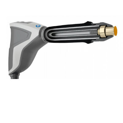
The Paris Radiology Institute (IRP; France) has installed nine Aixplorer ultrasound systems, which can acquire images 200 times faster than conventional systems.
The Paris Radiology Institute, founded in 1970, was the first private outpatient center in France to have a mammography scanner installed in 1986. Since 2006, the IRP has utilized two dedicated breast screening areas that feature three three-dimensional (3D) digital mammography units, one of which equipped for macro biopsies. The senography areas include eight dedicated ultrasound units, of which one is used for interventional mammary ultrasound.
The Aixplorer, a product of SuperSonic Imagine (Aix-en-Provence, France), can image two types of waves: ultrasound waves, which ensure excellent image quality; and shear waves, which allow physicians to visualize and analyze the stiffness of tissue in a real-time, reliable, reproducible, and noninvasive manner. Termed ShearWave Elastography, the technology significantly improves the detection, characterization, and monitoring of various pathologies involving the breast, liver, prostate, thyroid and others; it also reduces the number of needless biopsies.
“We have a team of 38 radiologists who excel in breast imaging. We were looking for an advanced ultrasound system providing high quality images and featuring breakthrough innovations to reinforce our senology ward,” declared the IRP in a press statement. “The Aixplorer by SuperSonic Imagine allows us to use ShearWave Elastography, which improves the precision of exams carried out by radiologists.”
“It is an honor for us to be chosen by the Paris Radiology Institute, a preeminent center that stays at the forefront of imaging technology and progress,” said Yves Tenaglia, vice president of SuperSonic Imagine Europe. “Our ultrasound system is not only easy to use, allowing radiologists to work comfortably and more productively, it is also highly advanced and innovative.”
Besides mammography, the IRP offer all types of imaging, including conventional radiology, ultrasound, vascular, computerized tomography (CT), magnetic resonance imaging (MRI), and bone density readings. The results of all tests can be reviewed and shared by doctors from any of seven units.
Ultrasonic shear-wave elastography is a form of vibrational wave analysis, similar to that of a seismograph during earthquakes. The main shockwave that propagates through the earth is a longitudinal wave, like that of ultrasound imaging, which runs along the direction of the wave. The secondary wave is a transverse wave that propagates by at right angles to the direction of the wave; these are also called shear waves or elastic shear waves. Shear waves are commonly used in nondestructive testing for flaws in manufactured materials, such as cracks.
Related Links:
Paris Radiology Institute
SuperSonic Imagine
Gold Member
STI Test
Vivalytic Sexually Transmitted Infection (STI) Array
Gold Member
POC Blood Gas Analyzer
Stat Profile Prime Plus
Silver Member
Wireless Mobile ECG Recorder
NR-1207-3/NR-1207-E
New
Radial Shock Wave Device
MASTERPULS »ultra«
Latest Hospital News News
- Nurse Tracking System Improves Hospital Workflow
- New Children’s Hospital Transforms California Healthcare
- Noisy Hospitals Face Threat of Decreased Federal Compensation
- Orthopedics Centre of Excellence Planned for Guy’s Hospital
- Research Suggests Avoidance of Low-Value Surgical Procedures
- U.S. Federal Readmission Fines Linked to Higher Mortality
- Columbia China to Build New Hospital in Jiaxing
- Dubai Debuts Second Robotic Pharmacy Service
- Seattle Hospital Network Shifts Away from Overlapping Surgeries
- ACC to Launch Valvular Heart Disease Program in China
- Mortality Rates Lower at Major Teaching Hospitals
- South Australia to Inaugurate Upscale Hospital
- Raffles to Launch Second Hospital Project in China
- Research Center Tackles Antimicrobial Drugs Challenge
- Miami Cardiac & Vascular Institute Completes Expansion Project
- Hospital Antibiotic Policies Improve Prescription Practices
Channels
Artificial Intelligence
view channel")
AI-Powered Algorithm to Revolutionize Detection of Atrial Fibrillation
Atrial fibrillation (AFib), a condition characterized by an irregular and often rapid heart rate, is linked to increased risks of stroke and heart failure. This is because the irregular heartbeat in AFib... Read more")
AI Diagnostic Tool Accurately Detects Valvular Disorders Often Missed by Doctors
Doctors generally use stethoscopes to listen for the characteristic lub-dub sounds made by heart valves opening and closing. They also listen for less prominent sounds that indicate problems with these valves.... Read more")
Critical Care
view channel")
Powerful AI Risk Assessment Tool Predicts Outcomes in Heart Failure Patients
Heart failure is a serious condition where the heart cannot pump sufficient blood to meet the body's needs, leading to symptoms like fatigue, weakness, and swelling in the legs and feet, and it can ultimately... Read more")
Peptide-Based Hydrogels Repair Damaged Organs and Tissues On-The-Spot
Scientists have ingeniously combined biomedical expertise with nature-inspired engineering to develop a jelly-like material that holds significant promise for immediate repairs to a wide variety of damaged... Read more")
One-Hour Endoscopic Procedure Could Eliminate Need for Insulin for Type 2 Diabetes
Over 37 million Americans are diagnosed with diabetes, and more than 90% of these cases are Type 2 diabetes. This form of diabetes is most commonly seen in individuals over 45, though an increasing number... Read moreSurgical Techniques
view channel")
Miniaturized Implantable Multi-Sensors Device to Monitor Vessels Health
Researchers have embarked on a project to develop a multi-sensing device that can be implanted into blood vessels like peripheral veins or arteries to monitor a range of bodily parameters and overall health status.... Read more")
Tiny Robots Made Out Of Carbon Could Conduct Colonoscopy, Pelvic Exam or Blood Test
Researchers at the University of Alberta (Edmonton, AB, Canada) are developing cutting-edge robots so tiny that they are invisible to the naked eye but are capable of traveling through the human body to... Read more")
Miniaturized Ultrasonic Scalpel Enables Faster and Safer Robotic-Assisted Surgery
Robot-assisted surgery (RAS) has gained significant popularity in recent years and is now extensively used across various surgical fields such as urology, gynecology, and cardiology. These surgeries, performed... Read morePatient Care
view channelFirst-Of-Its-Kind Portable Germicidal Light Technology Disinfects High-Touch Clinical Surfaces in Seconds
Reducing healthcare-acquired infections (HAIs) remains a pressing issue within global healthcare systems. In the United States alone, 1.7 million patients contract HAIs annually, leading to approximately... Read more")
Surgical Capacity Optimization Solution Helps Hospitals Boost OR Utilization
An innovative solution has the capability to transform surgical capacity utilization by targeting the root cause of surgical block time inefficiencies. Fujitsu Limited’s (Tokyo, Japan) Surgical Capacity... Read more")
Game-Changing Innovation in Surgical Instrument Sterilization Significantly Improves OR Throughput
A groundbreaking innovation enables hospitals to significantly improve instrument processing time and throughput in operating rooms (ORs) and sterile processing departments. Turbett Surgical, Inc.... Read more")
Health IT
view channel")
Machine Learning Model Improves Mortality Risk Prediction for Cardiac Surgery Patients
Machine learning algorithms have been deployed to create predictive models in various medical fields, with some demonstrating improved outcomes compared to their standard-of-care counterparts.... Read more")
Strategic Collaboration to Develop and Integrate Generative AI into Healthcare
Top industry experts have underscored the immediate requirement for healthcare systems and hospitals to respond to severe cost and margin pressures. Close to half of U.S. hospitals ended 2022 in the red... Read more")
AI-Enabled Operating Rooms Solution Helps Hospitals Maximize Utilization and Unlock Capacity
For healthcare organizations, optimizing operating room (OR) utilization during prime time hours is a complex challenge. Surgeons and clinics face difficulties in finding available slots for booking cases,... Read more")
AI Predicts Pancreatic Cancer Three Years before Diagnosis from Patients’ Medical Records
Screening for common cancers like breast, cervix, and prostate cancer relies on relatively simple and highly effective techniques, such as mammograms, Pap smears, and blood tests. These methods have revolutionized... Read morePoint of Care
view channel clearance for use with its Quantra QStat Cartridge (Photo courtesy of HemoSonics)")
Critical Bleeding Management System to Help Hospitals Further Standardize Viscoelastic Testing
Surgical procedures are often accompanied by significant blood loss and the subsequent high likelihood of the need for allogeneic blood transfusions. These transfusions, while critical, are linked to various... Read more prequalification (Photo courtesy of Cepheid)")
Point of Care HIV Test Enables Early Infection Diagnosis for Infants
Early diagnosis and initiation of treatment are crucial for the survival of infants infected with HIV (human immunodeficiency virus). Without treatment, approximately 50% of infants who acquire HIV during... Read more")
Whole Blood Rapid Test Aids Assessment of Concussion at Patient's Bedside
In the United States annually, approximately five million individuals seek emergency department care for traumatic brain injuries (TBIs), yet over half of those suspecting a concussion may never get it checked.... Read more")
New Generation Glucose Hospital Meter System Ensures Accurate, Interference-Free and Safe Use
A new generation glucose hospital meter system now comes with several features that make hospital glucose testing easier and more secure while continuing to offer accuracy, freedom from interference, and... Read moreBusiness
view channel")
Johnson & Johnson Acquires Cardiovascular Medical Device Company Shockwave Medical
Johnson & Johnson (New Brunswick, N.J., USA) and Shockwave Medical (Santa Clara, CA, USA) have entered into a definitive agreement under which Johnson & Johnson will acquire all of Shockwave’s... Read more")
")