Interactive VR Assists in Treatment Planning
|
By HospiMedica International staff writers Posted on 16 Apr 2018 |

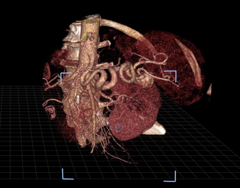
Image: An image of a splenic artery aneurysm as seen in virtual reality (Photo courtesy of SIR).
Interactive virtual reality (VR) shows a patient's unique internal anatomy to interventional radiologists in order to help physicians effectively prepare and tailor their approach to complex treatments, such as splenic artery aneurysm repair. In a new study, researchers compared the VR technology to the use of images from a commonly used visualization software system that displays images on a standard two-dimensional platform and found the accuracy to be similar with both methods, though confidence improved substantially with VR.
VR converts a patient's pre-procedural CT scans into 3D images that can be virtually moved and examined by radiologists wearing virtual reality-type glasses. VR allows operators to manipulate routine, two-dimensional images in an open three-dimensional space in order to allow them to look into the patients' organs and tissues that had been impossible outside of the human body, until now. This arms the operators with a deeper and intuitive understanding of spatial relationships, such as between an aneurysm and the surrounding arteries.
In the study, three radiologists used both the technologies to independently evaluate 17 splenic artery aneurysms in 14 patients. The researchers measured the accuracy of the radiologists in identifying inflow and outflow arteries associated with the aneurysms with each method. The radiologists also ranked improvements in their confidence on a four-point scale when using VR as compared to the standard method. The researchers found that 93% of the participating physicians who used the VR method indicated higher confidence in their abilities (a score of at least three).
"Treating splenic artery aneurysms can be very difficult because of their intricate nature and anatomic variations from patient to patient. This new platform allows you to view a patient's arterial anatomy in a three-dimensional image, as if it is right in front of you, which may help interventional radiologists more quickly and thoroughly plan for the equipment and tools they'll need for a successful outcome," said Zlatko Devcic, M.D., a fellow of interventional radiology at Stanford University School of Medicine and collaborating author of the study. "Pre-operative planning is possibly the most important step towards successfully treating a patient, so the value of VR cannot be understated," Devcic said. "This technology gives us a totally different way to look at that structure and safely plan our approach to patient care."
The research was presented at the Society of Interventional Radiology's 2018 Annual Scientific Meeting. The researchers hope that more studies in the future will examine whether the VR technology will ultimately help reduce the time required to perform the treatment and consequently, reduce the amount of radiation and contrast exposure to the patient.
Latest Business News
- Johnson & Johnson Acquires Cardiovascular Medical Device Company Shockwave Medical
- Mindray to Acquire Chinese Medical Device Company APT Medical
- Olympus Acquires Korean GI Stent Maker Taewoong Medical
- Karl Storz Acquires British AI Specialist Innersight Labs
- Stryker to Acquire French Joint Replacement Company SERF SAS
- Medical Illumination Acquires Surgical Lighting Specialist Isolux
- 5G Remote-Controlled Robots to Enable Even Cross-Border Surgeries

- International Hospital Federation Announces 2023 IHF Award Winners
- Unprecedented AI Integration Transforming Surgery Landscape, Say Experts

- New WHO Guidelines to Revolutionize AI in Healthcare
- Getinge Acquires US-Based Medical Equipment Provider Healthmark Industries
- Global Surgical Lights Market Driven by Increasing Number of Procedures
- Global Capsule Endoscopy Market Driven by Demand for Accurate Diagnosis of Gastrointestinal Conditions
- Global OR Integration Market Driven by Need for Improved Workflow Efficiency and Productivity
- Global Endoscopy Devices Market Driven by Increasing Adoption of Endoscopes in Surgical Procedures
- Global Minimally Invasive Medical Devices Market Driven by Benefits of MIS Procedures
Channels
Artificial Intelligence
view channel
AI-Powered Algorithm to Revolutionize Detection of Atrial Fibrillation
Atrial fibrillation (AFib), a condition characterized by an irregular and often rapid heart rate, is linked to increased risks of stroke and heart failure. This is because the irregular heartbeat in AFib... Read more
AI Diagnostic Tool Accurately Detects Valvular Disorders Often Missed by Doctors
Doctors generally use stethoscopes to listen for the characteristic lub-dub sounds made by heart valves opening and closing. They also listen for less prominent sounds that indicate problems with these valves.... Read moreCritical Care
view channel
Powerful AI Risk Assessment Tool Predicts Outcomes in Heart Failure Patients
Heart failure is a serious condition where the heart cannot pump sufficient blood to meet the body's needs, leading to symptoms like fatigue, weakness, and swelling in the legs and feet, and it can ultimately... Read more
Peptide-Based Hydrogels Repair Damaged Organs and Tissues On-The-Spot
Scientists have ingeniously combined biomedical expertise with nature-inspired engineering to develop a jelly-like material that holds significant promise for immediate repairs to a wide variety of damaged... Read more
One-Hour Endoscopic Procedure Could Eliminate Need for Insulin for Type 2 Diabetes
Over 37 million Americans are diagnosed with diabetes, and more than 90% of these cases are Type 2 diabetes. This form of diabetes is most commonly seen in individuals over 45, though an increasing number... Read moreSurgical Techniques
view channel
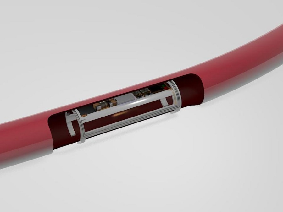
Miniaturized Implantable Multi-Sensors Device to Monitor Vessels Health
Researchers have embarked on a project to develop a multi-sensing device that can be implanted into blood vessels like peripheral veins or arteries to monitor a range of bodily parameters and overall health status.... Read more
Tiny Robots Made Out Of Carbon Could Conduct Colonoscopy, Pelvic Exam or Blood Test
Researchers at the University of Alberta (Edmonton, AB, Canada) are developing cutting-edge robots so tiny that they are invisible to the naked eye but are capable of traveling through the human body to... Read more
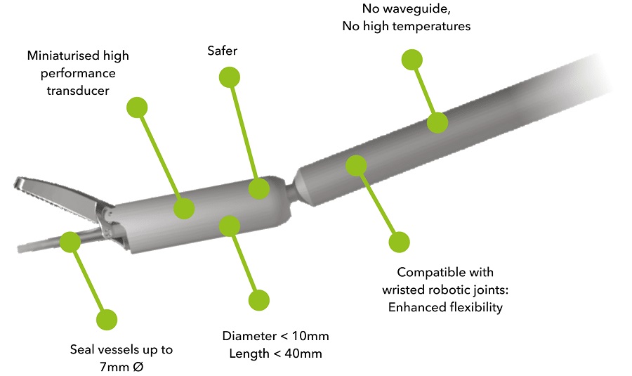
Miniaturized Ultrasonic Scalpel Enables Faster and Safer Robotic-Assisted Surgery
Robot-assisted surgery (RAS) has gained significant popularity in recent years and is now extensively used across various surgical fields such as urology, gynecology, and cardiology. These surgeries, performed... Read morePatient Care
view channelFirst-Of-Its-Kind Portable Germicidal Light Technology Disinfects High-Touch Clinical Surfaces in Seconds
Reducing healthcare-acquired infections (HAIs) remains a pressing issue within global healthcare systems. In the United States alone, 1.7 million patients contract HAIs annually, leading to approximately... Read more
Surgical Capacity Optimization Solution Helps Hospitals Boost OR Utilization
An innovative solution has the capability to transform surgical capacity utilization by targeting the root cause of surgical block time inefficiencies. Fujitsu Limited’s (Tokyo, Japan) Surgical Capacity... Read more
Game-Changing Innovation in Surgical Instrument Sterilization Significantly Improves OR Throughput
A groundbreaking innovation enables hospitals to significantly improve instrument processing time and throughput in operating rooms (ORs) and sterile processing departments. Turbett Surgical, Inc.... Read moreHealth IT
view channel
Machine Learning Model Improves Mortality Risk Prediction for Cardiac Surgery Patients
Machine learning algorithms have been deployed to create predictive models in various medical fields, with some demonstrating improved outcomes compared to their standard-of-care counterparts.... Read more
Strategic Collaboration to Develop and Integrate Generative AI into Healthcare
Top industry experts have underscored the immediate requirement for healthcare systems and hospitals to respond to severe cost and margin pressures. Close to half of U.S. hospitals ended 2022 in the red... Read more
AI-Enabled Operating Rooms Solution Helps Hospitals Maximize Utilization and Unlock Capacity
For healthcare organizations, optimizing operating room (OR) utilization during prime time hours is a complex challenge. Surgeons and clinics face difficulties in finding available slots for booking cases,... Read more
AI Predicts Pancreatic Cancer Three Years before Diagnosis from Patients’ Medical Records
Screening for common cancers like breast, cervix, and prostate cancer relies on relatively simple and highly effective techniques, such as mammograms, Pap smears, and blood tests. These methods have revolutionized... Read morePoint of Care
view channel
Critical Bleeding Management System to Help Hospitals Further Standardize Viscoelastic Testing
Surgical procedures are often accompanied by significant blood loss and the subsequent high likelihood of the need for allogeneic blood transfusions. These transfusions, while critical, are linked to various... Read more
Point of Care HIV Test Enables Early Infection Diagnosis for Infants
Early diagnosis and initiation of treatment are crucial for the survival of infants infected with HIV (human immunodeficiency virus). Without treatment, approximately 50% of infants who acquire HIV during... Read more
Whole Blood Rapid Test Aids Assessment of Concussion at Patient's Bedside
In the United States annually, approximately five million individuals seek emergency department care for traumatic brain injuries (TBIs), yet over half of those suspecting a concussion may never get it checked.... Read more


